Volume 10, Number 12—December 2004
Research
Nonsusceptibility of Primate Cells to Taura Syndrome Virus
Figure 4
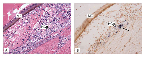
Figure 4. Covert Taura syndrome virus (TSV) infection (transition/chronic phase of TS disease) in indicator specific pathogen free–Litopenaeus vannamei shrimp was confirmed by in situ hybridization (ISH) with digoxigenin-labeled gene probes specific for detection of TSV. A) Histologic section through the dorsal cuticular epithelium showing a melanized resolving lesion (MZ) and hemolytic congestion (HCg), indicative of the transition phase of TSV infection (hematoxylin/eosin-phloxin stain; 50x) . B) TSV ISH on the consecutive section to that shown in 5A, where binding of the TSV probes is shown by the black precipitate (arrow) indicating the presence of TSV within the cytoplasm of cells of the cuticular epithelium (Bismarck Brown counterstain; 50x).