Volume 11, Number 12—December 2005
Dispatch
Human Rickettsia felis Infection, Canary Islands, Spain
Figure
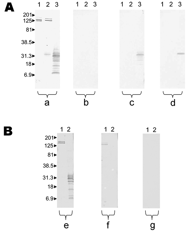
Figure. Results of Western blot performed with serum samples from patient 5 with Rickettsia felis infection and patient 10 with R. typhi infection. Molecular masses (in kilodaltons) are given to the left of panels. A) Patient with R. felis infection; a, untreated serum analyzed by using R. conorii (lane 1), R. typhi (lane 2), and R. felis (lane 3); b, R. felis–adsorbed serum analyzed by using R. conorii (lane 1), R. typhi (lane 2), R. felis (lane 3); all antibodies were removed; c, R. typhi–adsorbed serum analyzed by using R. typhi (lane 1) and R. felis (lane 2); antibodies to R. felis remained; d, R. conorii–adsorbed serum analyzed by using R. conorii (lane 1), R. typhi (lane 2), R. felis (lane 3); antibodies to R. felis remained. B) Patient with murine typhus; e, untreated serum analyzed by using R. typhi (lane 1) and R. felis (lane 2); f, R. felis–adsorbed serum analyzed by using R. typhi (lane 1) and R. felis (lane 2); antibodies to R. typhi remained; g, R. typhi–adsorbed serum analyzed by using R. typhi (lane 1) and R. felis (lane 2); all antibodies were removed.
1These authors contributed equally to this article.