Volume 14, Number 4—April 2008
Letter
Rickettsia sibirica subsp. mongolitimonae Infection and Retinal Vasculitis
To the Editor: Rickettsia sibirica subsp. mongolitimonae is an intracellular bacterium that belongs to the species R. sibirica (1). To date, only 11 cases of infection with this bacterium have been reported (2–6). We report a case in a pregnant woman with ocular vasculitis.
A 20-year-old woman in the 10th week of her pregnancy was admitted in June 2005 to St. Eloi Hospital in Montpellier, France, with an 8-day history of fever, eschar, hemifacial edema, and headache. On examination the day of admission, she had a fever of 38.5°C, headache, and frontal eschar surrounded by an inflammatory halo. Painful retroauricular and cervical lymphadenopathies were noted. Results of a clinical examination were otherwise within normal limits. No tick bite was reported by the patient, although she had been walking a few days before in Camargue (southern France). Serologic results for R. conorii, R. typhi, Brucella spp., Borrelia spp., and Coxiella burnetii were negative.
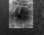
One day after admission, she reported loss of vision (scotoma) in her right eye. She underwent a complete ophthalmic evaluation. Measurement of visual acuity and results of a slit-lamp examination were within normal limits, but a funduscopic examination showed a white retinal macular lesion that corresponded in a fluorescein angiograph to an area of retinal ischemia induced by vascular inflammation and subsequent occlusion (Figure). The following day, a rash with a few maculopapular elements developed, which involved only the palms of the hands and soles of the feet. Mediterranean spotted fever was suspected. Cyclines and fluoroquinolones were contraindicated because of her pregnancy, and the patient had a history of maculopapular rash after taking josamycin. She was treated with azithromycin, 500 mg/day for 10 days, under close surveillance. After 2 days of treatment, she was afebrile and the rash completely resolved. No obstetric complications occurred and she gave birth to a healthy boy at term. Two years later, the right scotoma remained unchanged.
Serologic tests for rickettsiosis were performed with an acute-phase serum sample and a convalescent-phase serum sample (1 month after onset of symptoms). Samples were sent to the World Health Organization Collaborative Center in Marseille for rickettsial reference and research. Immunoglobulin (Ig) G and IgM titers were estimated by using a microimmunofluorescence assay; results were negative. Culture of a skin biopsy specimen from the eschar showed negative results.
DNA was extracted from eschar biopsy specimen and used as template in a PCR with primers complementary to portions of the coding sequences of the rickettsial outer membrane protein A and citrate synthase genes as described (5). Nucleotide sequences of the PCR products were determined. All sequences shared 100% similarity with R. sibirica subsp. mongolotimonae when compared with those in the GenBank database.
Infections caused by R. sibirica subsp. mongolitimonae have been reported as lymphangitis-associated rickettsiosis (4). Our case-patient had the clinical symptoms reported for this disease: fever, maculopapular rash, eschar, enlarged satellite lymph nodes, and lymphangitis. Seasonal occurrence of this disease in the spring is common and has been reported in 9 of 12 cases, including the case-patient reported here (2–6). A total of 75% of these R. sibirica subsp. mongolitimonae infections occurred in southern France; other cases have been recently reported in Greece (5), Portugal (6), and South Africa (7). However, the vector of R. sibirica subsp. mongolitimonae has not been identified (7). This rickettsia has been isolated from Hyalomma asiaticum ticks in Inner Mongolia, from H. truncatum in Niger (8), and from H. anatolicum excavatum in Greece (5). Hyalomma spp. ticks are suspected of being the vector and are widespread in Africa, southeastern Europe (including France), and Asia.
Rickettsiosis caused by R. rickettsii and R. conorii during pregnancy has been reported without risk for vertical transmission (9). First-line antimicrobial drugs used to treat rickettsial disease are cyclines and quinolones, but they are contraindicated during pregnancy. Chloramphenicol is an alternative drug for pregnant women but it is not available in France. Macrolides (azithromycin, clarithromycin, and josamycin) are effective against rickettsial disease and can be used safely during pregnancy.
No ocular complications were reported in the 11 previous cases of rickettsiosis caused by R. subsp. mongolitimonae. However, ocular lesions, including optic disk staining, white retinal lesions, retinal hemorrhages, multiple hypofluorescent choroidal dots, mild vitritis, and retinal vasculitis, have been described in patients with rickettsiosis caused by R. conorii, R. rickettsii, and R. typhi (10). Most of these posterior segment manifestations are usually asymptomatic in patients with acute Mediterranean spotted fever (10) and can be easily overlooked. Retinal vasculitis was reported in 45%–55% of the patients, but retinal artery occlusion secondary to vasculitis has been described in only 2 cases of infection with R. conorii and R. rickettsii (10) without details of clinical symptoms. Because ocular involvement could be asymptomatic and easily overlooked, an ophthalmic evaluation should be conducted when rickettsiosis is suspected.
References
- Fournier PE, Zhu Y, Yu X, Raoult D. Proposal to create subspecies of Rickettsia sibirica and an amended description of Rickettsia sibirica. Ann N Y Acad Sci. 2006;1078:597–606. DOIPubMedGoogle Scholar
- Raoult D, Brouqui P, Roux V. A new spotted-fever-group rickettsiosis. Lancet. 1996;348:412. DOIPubMedGoogle Scholar
- Fournier PE, Tissot-Dupont H, Gallais H, Raoult D. Rickettsia mongolotimonae: a rare pathogen in France. Emerg Infect Dis. 2000;6:290–2.PubMedGoogle Scholar
- Fournier PE, Gouriet F, Brouqui P, Lucht F, Raoult D. Lymphangitis-associated rickettsiosis, a new rickettsiosis caused by Rickettsia sibirica mongolotimonae: seven new cases and review of the literature. Clin Infect Dis. 2005;40:1435–44. DOIPubMedGoogle Scholar
- Psaroulaki A, Germanakis A, Gikas A, Scoulica E, Tselentis Y. Simultaneous detection of “Rickettsia mongolotimonae” in a patient and in a tick in Greece. J Clin Microbiol. 2005;43:3558–9. DOIPubMedGoogle Scholar
- de Sousa R, Barata C, Vitorino L, Santos-Silva M, Carrapato C, Torgal J, Rickettsia sibirica isolation from a patient and detection in ticks, Portugal. Emerg Infect Dis. 2006;12:1103–8.PubMedGoogle Scholar
- Pretorius AM, Birtles RJ. Rickettsia mongolotimonae infection in South Africa. Emerg Infect Dis. 2004;10:125–6.PubMedGoogle Scholar
- Parola P, Inokuma H, Camicas JL, Brouqui P, Raoult D. Detection and identification of spotted fever group rickettsiae and ehrlichiae in African ticks. Emerg Infect Dis. 2001;7:1014–7.PubMedGoogle Scholar
- Stallings SP. Rocky Mountain spotted fever and pregnancy: a case report and review of the literature. Obstet Gynecol Surv. 2001;56:37–42. DOIPubMedGoogle Scholar
- Khairallah M, Ladjimi A, Chakroun M, Messaoud R, Yahia SB, Zaouali S, Posterior segment manifestations of Rickettsia conorii infection. Ophthalmology. 2004;111:529–34. DOIPubMedGoogle Scholar
Figure
Cite This ArticleRelated Links
Table of Contents – Volume 14, Number 4—April 2008
| EID Search Options |
|---|
|
|
|
|
|
|

Please use the form below to submit correspondence to the authors or contact them at the following address:
Didier Bessis, Service de Dermatologie, Hôpital Saint-Eloi, 80 Ave Augustin-Fliche, 34295 Montpellier CEDEX 5, France;
Top