Volume 16, Number 7—July 2010
Dispatch
Cryptococcus gattii Genotype VGIIa Infection in Man, Japan, 2007
Koh Okamoto, Shuji Hatakeyama , Satoru Itoyama, Yoko Nukui, Yusuke Yoshino, Takatoshi Kitazawa, Hiroshi Yotsuyanagi, Reiko Ikeda, Takashi Sugita, and Kazuhiko Koike
Figure
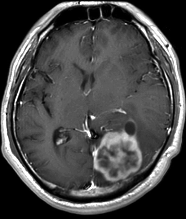
Figure. Postcontrast T1-weighted magnetic resononance image of the brain of a 44-year-old man with cerebral cryptococomma in Japan, 2007, showing a rim-enhancing lobulated mass (lower right) with surrounding edema in the left occipital lobe.
Page created: March 02, 2011
Page updated: March 02, 2011
Page reviewed: March 02, 2011
The conclusions, findings, and opinions expressed by authors contributing to this journal do not necessarily reflect the official position of the U.S. Department of Health and Human Services, the Public Health Service, the Centers for Disease Control and Prevention, or the authors' affiliated institutions. Use of trade names is for identification only and does not imply endorsement by any of the groups named above.