Volume 20, Number 8—August 2014
Letter
Diagnosis of Bartonella henselae Prosthetic Valve Endocarditis in Man, France
To the Editor: Bartonella spp. cause 2% of cases of blood culture–negative endocarditis (1). Early diagnosis of Bartonella spp. infectious endocarditis, is challenging, especially for patients with preexisting valvular heart disease. A diagnosis for these patients requires bacterial culture, serologic testing, or molecular detection in serum or tissue (2). The sensitivity and specificity of Duke modified criteria (3) for detecting endocardial involvement by echocardiography are not optimal, which results in decreased diagnostic accuracy (4).
18Fluorodeoxyglucose-positron emission tomography/computed tomography (18FDG-PET/CT), has been shown to be beneficial for diagnosis (4) and management of prosthetic valve endocarditis (5), particularly if echocardiographic findings are inconclusive (6). This procedure can be performed in patients of all ages by adjusting the dose of 18FDG to the weight of the patient. We report a case that illustrates the usefulness of 18FDG-PET/CT for diagnosis of Bartonella henselae infectious endocarditis in a patient with a prosthetic valve.
On October 18, 2012, a 56-year-old man was admitted to Timone Hospital (Marseille, France) with fatigue and weight loss (–6 kg) over the past 6 months. He had had an aortic valve replacement and a bioprosthesis was inserted in 2005 for rheumatic disease. The patient had owned a kitten for 6 months.
Laboratory findings showed moderate anemia (hemoglobin level 114 g/L), an elevated C-reactive protein level (34.5 mg/L), and polyclonal hypergammaglobulinemia. A test result was negative for rheumatoid factor. Transthoracic and transesophageal echocardiograms showed a thickened and partial aortic stenosis around the bioprosthesis.
Because infectious endocarditis was suspected, treatment with intravenous antimicrobial drugs (amoxicillin, 200 mg/kg/day for 6 weeks and gentamicin, 160 mg/day for 2 weeks) was initiated. On day 7, he was transferred to the cardiology department of Timone Hospital in Marseille, France. An endocarditis test was performed by using an endocarditis kit as described (7). Three routine blood cultures were negative. The patient was given a diagnosis of possible infectious endocarditis by using the Duke score (3).
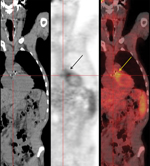
An 18FDG-PET/CT scan was performed and showed 18FDG uptake in the aortic bioprosthesis area (Figure). Results of a whole body scan were normal. An immunofluorescence test for Bartonella spp. showed titers of 400 for IgG against B. quintana and B. henselae (1), and Western blot confirmed a reactivity pattern pathognomonic for B. henselae endocarditis. Results of a PCR performed with a blood sample stored in EDTA (1) were positive for B. henselae. On day 13, antimicrobial drug therapy was changed to oral doxycycline, 200 mg/day for 1 month and intravenous gentamicin, 160 mg/day for 15 days. On day 28, the patient was released from the hospital.
Two weeks later while still taking doxycycline, the patient was readmitted for a subarachnoid hemorrhage caused by a ruptured cerebral mycotic aneurysm. After arterial ligation, intravenous gentamicin was given with doxycycline for an additional 15 days. The patient recovered during this therapy.
On February 25, 2013, because of valvular stenosis, the patient underwent a new replacement with an aortic bioprosthesis. Valve culture remained sterile, but a PCR result was positive for B. henselae. Histologic analysis of the valve with Warthin-Starry stain showed no microorganisms. The patient remained asymptomatic for 2 months after surgery.
Diagnosis of possible infectious endocarditis in the patient was suspected after results of a transesophageal echocardiogram were used as a major criterion (thickened and partial aortic stenosis), and predisposing heart condition (aortic bioprosthesis) were used as a minor criterion (3) Because blood cultures were negative for the organism, the diagnosis of B. henselae infection was made by using serologic analysis and PCR (7). 18FDG-PET/CT was especially valuable in early diagnosis of infectious endocarditis because echocardiography showed no vegetation or abscess, a common feature of Bartonella spp. endocarditis in which vegetations are often small or absent. In addition, a diagnosis of infectious endocarditis remains challenging, particularly in cases of prosthetic valve infections, in which results of initial echocardiography are not useful in 30% of cases (5). In such cases, the diagnostic accuracy of modified Duke criteria decreases.
Use of 18FDG-PET/CT for diagnosis and monitoring of infectious endocarditis showed promising results, particularly for prosthetic valve infections (5), cardiac device–related infections (6), and when results of echocardiography are inconclusive or blood cultures are negative (5). 18FDG-PET/CT with abnormal uptake of FDG was proposed as a diagnostic criterion of prosthetic valve infectious endocarditis (4). In contrast, a negative 18FDG-PET/CT result does not rule out infectious endocarditis (8).
Use of 18FDG-PET/CT has been discussed mainly because of problems of sensitivity of tracer uptake in heart tissue and small vegetations (9). Improvements, such as patient preparation with low carbohydrate–fat diet and technical advances in the newest 18FDG-PET/CT scanners, may increase this sensitivity in future studies (10). Although 18FDG-PET/CT will not replace clinical evaluation, laboratory tests, and echocardiography, this procedure might be helpful in diagnosis of Bartonella spp. infectious endocarditis.
Acknowledgment
We thank Clémence Bassez and Laurie-Anne Mansou for patient management.
References
- Brouqui P, Raoult D. Endocarditis due to rare and fastidious bacteria. Clin Microbiol Rev. 2001;14:177–207. DOIPubMedGoogle Scholar
- Fournier PE, Lelievre H, Eykyn SJ, Mainardi JL, Marrie TJ, Bruneel F, Epidemiologic and clinical characteristics of Bartonella quintana and Bartonella henselae endocarditis: a study of 48 patients. Medicine (Baltimore). 2001;80:245–51. DOIPubMedGoogle Scholar
- Li JS, Sexton DJ, Mick N, Nettles R, Fowler VG Jr, Ryan T, Proposed modifications to the Duke criteria for the diagnosis of infective endocarditis. Clin Infect Dis. 2000;30:633–8. DOIPubMedGoogle Scholar
- Basu S, Chryssikos T, Moghadam-Kia S, Zhuang H, Torigian DA, Alavi A. Positron emission tomography as a diagnostic tool in infection: present role and future possibilities. Semin Nucl Med. 2009;39:36–51. DOIPubMedGoogle Scholar
- Saby L, Laas O, Habib G, Cammilleri S, Mancini J, Tessonnier L, Positron emission tomography/computed tomography for diagnosis of prosthetic valve endocarditis: increased valvular 18F-fluorodeoxyglucose uptake as a novel major criterion. J Am Coll Cardiol. 2013;61:2374–82. DOIPubMedGoogle Scholar
- Millar BC, Prendergast BD, Alavi A, Moore JE. (18)FDG-positron emission tomography (PET) has a role to play in the diagnosis and therapy of infective endocarditis and cardiac device infection. Int J Cardiol. 2013;167:1724–36. DOIPubMedGoogle Scholar
- Raoult D, Casalta JP, Richet H, Khan M, Bernit E, Rovery C, Contribution of systematic serological testing in diagnosis of infective endocarditis. J Clin Microbiol. 2005;43:5238–42. DOIPubMedGoogle Scholar
- Sankatsing SU, Kolader ME, Bouma BJ, Bennink RJ, Verberne HJ, Ansink TM, 18F-fluoro-2-deoxyglucose positron emission tomography-negative endocarditis lenta caused by Bartonella henselae. J Heart Valve Dis. 2011;20:100–2 .PubMedGoogle Scholar
- Kouijzer IJ, Vos FJ, Janssen MJ, van Dijk AP, Oyen WJ, Bleeker-Rovers CP. The value of 18FDG PET/CT in diagnosing infectious endocarditis. Eur J Nucl Med Mol Imaging. 2013;40:1102–7. DOIPubMedGoogle Scholar
- Williams G, Kolodny GM. Suppression of myocardial 18F-FDG uptake by preparing patients with a high-fat, low-carbohydrate diet. AJR Am J Roentgenol. 2008;190:W151–6. DOIPubMedGoogle Scholar
Figure
Cite This ArticleRelated Links
Table of Contents – Volume 20, Number 8—August 2014
| EID Search Options |
|---|
|
|
|
|
|
|

Please use the form below to submit correspondence to the authors or contact them at the following address:
Didier Raoult, Aix Marseille Université, URMITE, UM 63, CNRS 7278, IRD 198, INSEM U1095, 13005 Marseille, France
Top