Volume 22, Number 7—July 2016
Letter
Porcine Bocavirus Infection Associated with Encephalomyelitis in a Pig, Germany1
Figure
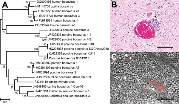
Figure. Phylogenetic analysis and staining of porcine bocavirus (PBoV) from the spinal cord of a diseased pig, Hannover, Germany. A) Phylogenetic relationship of PBoV isolate S1142/13 (bold) with other bocaviruses. The nucleotide sequence of the nearly complete viral protein 1 of PBoV S1142/13 was aligned with other members of the genus Bocaparvovirus, and a maximum-likelihood phylogenetic tree was prepared by using the general time reversible plus invariable sites plus gamma distribution model and 500 bootstrap replicates. Only bootstrap values >70 are shown. Scale bar indicates nucleotide substitutions per site. B) Spinal cord of the diseased pig showing perivascular accentuated mild, focal, nonsuppurative inflammation (arrow). Hematoxylin and eosin stain. Scale bar indicates 100 µm. C) Intracytoplasmic and intranuclear PBoV-specific red positive signals in neurons of the spinal cord of the diseased pig (arrowheads), determined by using fluorescent in situ hybridization (Fast Red; ViewRNA Chromogenic Signal Amplification Kit; Affymetrix, Santa Clara, CA, USA). Phase contrast and fluorescent microscopy. Scale bar indicates 100 µm.
1Preliminary results from this study were presented at the 3rd International One Health Congress, March 15–18, 2015, Amsterdam, the Netherlands, and at the Conference of the German Veterinary Medical Association, March 8–10, 2015, Fulda, Germany.