Volume 23, Number 2—February 2017
Dispatch
Fatal Infection with Murray Valley Encephalitis Virus Imported from Australia to Canada, 2011
Figure 2
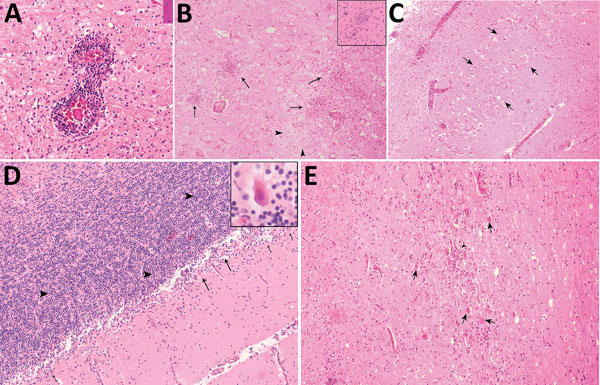
Figure 2. Hematoxylin and eosin–stained autopsy specimens from a patient with a fatal infection of Murray Valley encephalitis virus imported from Australia to Canada, 2011. A) Pons showing perivascular inflammatory infiltrate (original magnification ×40). B) Thalamus showing extensive inflammation (arrows) surrounding an area of rarefaction caused by necrosis (arrowheads) and neuronal loss (original magnification ×10); inset shows a microglial nodule (original magnification ×20). C) Pyramidal cell layer of the hippocampus showing extensive acute neuronal death (arrows) (original magnification ×4). D) Cerebellum showing severe depletion of Purkinje neurons and acute neuronal death (arrows and inset [original magnification ×40]) with relative sparing of the internal granule cell layer (arrowheads) and inflammation (short arrows) (original magnification ×10). E) Substantia nigra showing extensive inflammation, acute neuronal death (arrows), neuronophagia (arrowhead), and gliosis (original magnification ×10).