Volume 24, Number 1—January 2018
Research Letter
Inonotosis in Patient with Hematologic Malignancy
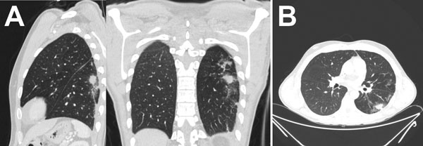
Figure 1

Figure 1. Computed tomography of the lungs in patient with invasive fungal disease caused by Inonotus spp., Madrid, Spain. Images show pulmonary nodule with halo sign in the left superior lobe, with peripheral distribution.
1These authors contributed equally to this article.
Page created: December 19, 2017
Page updated: December 19, 2017
Page reviewed: December 19, 2017
The conclusions, findings, and opinions expressed by authors contributing to this journal do not necessarily reflect the official position of the U.S. Department of Health and Human Services, the Public Health Service, the Centers for Disease Control and Prevention, or the authors' affiliated institutions. Use of trade names is for identification only and does not imply endorsement by any of the groups named above.