Mycobacterium marseillense Infection in Human Skin, China, 2018
Bibo Xie
1, Yanqing Chen
1, Jian Wang, Wei Gao, Haiqing Jiang, Jiya Sun, Xindong Jin, Xudong Sang, Xiaobing Yu, and Hongsheng Wang

Author affiliations: Institute of Dermatology, Chinese Academy of Medical Sciences and Peking Union Medical College, Nanjing, China (B. Xie,Y. Chen, W. Gao, H. Jiang, H. Wang); Zhejiang Institute of Dermatology, Deqing, China (B. Xie, J. Wang, X. Jin, X. Sang, X. Yu); Jiangsu Key Laboratory of Molecular Biology for Skin Diseases and STIs, Nanjing (Y. Chen, W. Gao, H. Jiang, H. Wang); Suzhou Institute of Systems Medicine, Chinese Academy of Medical Sciences, Suzhou, China (J. Sun); Centre for Global Health, Nanjing Medical University, Nanjing (H. Wang)
Main Article
Figure

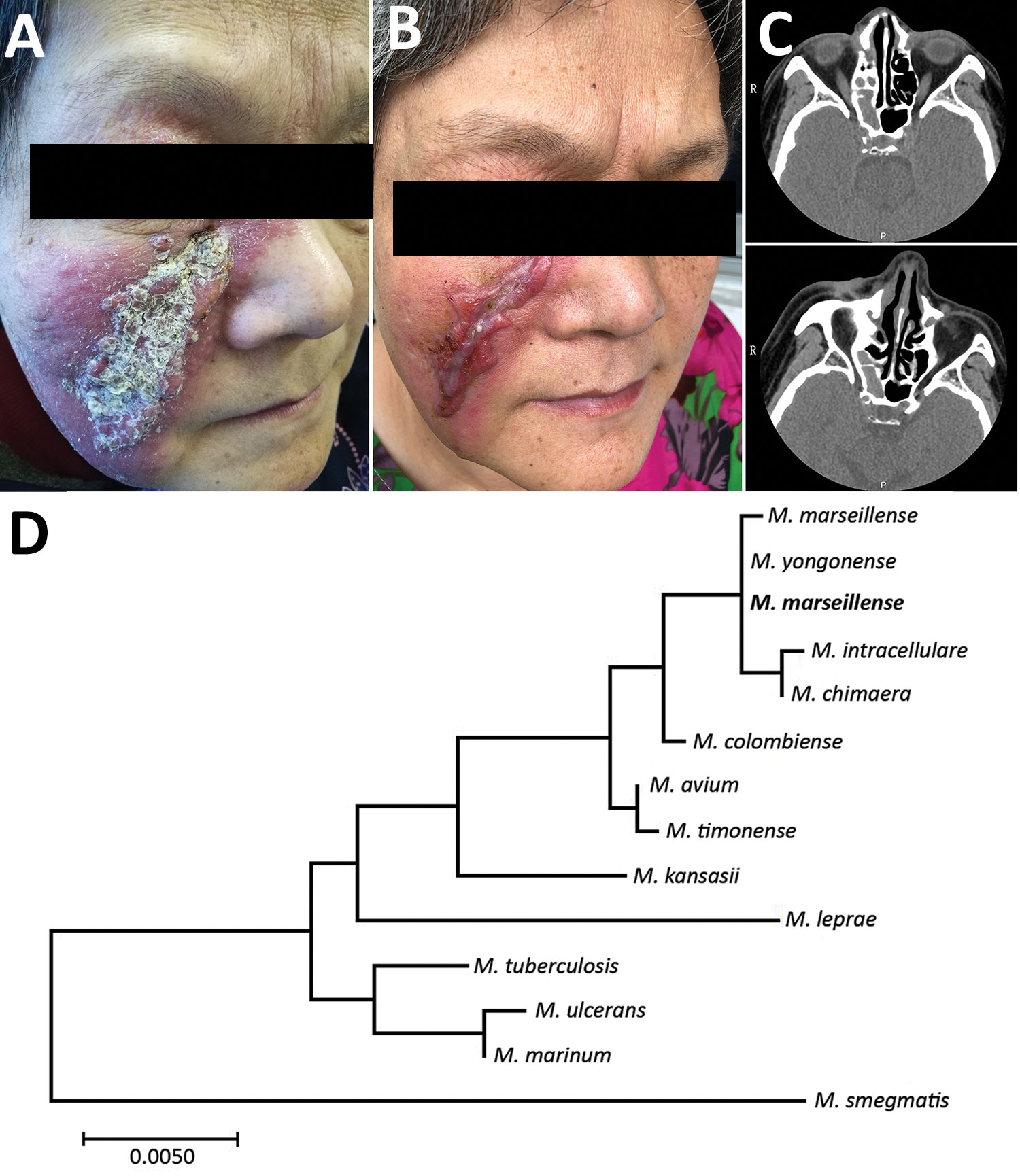
Figure. Skin lesions and computer tomography scans of woman with Mycobacterium marseillense skin infection, China, 2018, and genomic analysis of isolate. A, B) Facial skin lesion of woman with M. marseillense infection before and after treatment. Infiltrated erythematous plaque with yellowish scales and crusts (A) resolved to a scar after clearance of infection (B). C) Computed tomography imaging before treatment (top) shows heterogeneous hypersignal in right ethmoid sinus and after treatment (bottom) shows recovery of right ethmoid sinus. P, posterior; R, right. D) Phylogenetic tree constructed with 16S rRNA gene sequence of isolate from patient (bold) and other species. Scale bar indicates nucleotide substitutions per site.
Main Article
Page created: September 17, 2019
Page updated: September 17, 2019
Page reviewed: September 17, 2019
The conclusions, findings, and opinions expressed by authors contributing to this journal do not necessarily reflect the official position of the U.S. Department of Health and Human Services, the Public Health Service, the Centers for Disease Control and Prevention, or the authors' affiliated institutions. Use of trade names is for identification only and does not imply endorsement by any of the groups named above.
