Volume 26, Number 10—October 2020
Dispatch
Polyester Vascular Graft Material and Risk for Intracavitary Thoracic Vascular Graft Infection1
Figure
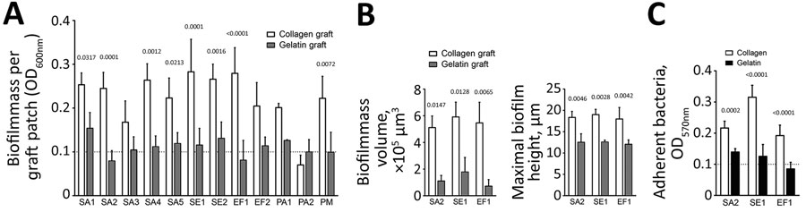
Figure. Increased susceptibility of collagen graft to biofilm formation compared with gelatin graft. Graft patches were inoculated with indicated bacterial strains for 72 h and analyzed quantitatively and qualitatively. A) Biofilm formation on the graft patches determined by optical density measurements. B) Total biofilm mass volume and maximal biofilm height, respectively, formed on the graft patches by the 3 clinical isolates—SA2, SE1, and EF— determined from the confocal laser scanning microscopy images with imaris software (https://imaris.oxinst.com). C) Adherence assay to the 2 different coatings used by the grafts. The limit of reliable detection of the plate reader is indicated by the dashed line ( OD600nm = 0.1). All data represent mean ± SD of 3 biological replicates performed in at least 2 technical replicates and were analyzed by using GraphPad Prism 8 (GraphPad Software, https://www.graphpad.com). The values above the graphs represent p values, calculated by using 2-way analysis of variance with the Sidak multiple comparison to determine statistical significance between the 2 graft types or coatings (panels A–C). EF, Enterococcus faecalis; OD600nm, optical density at a wavelength of 600; PA, Pseudomonas aeruginosa; PM, Pasteurella multocida; PVGI, prosthetic vascular graft infection; SA, Staphylococcus aureus; SE, S. epidermidis.
1This work was presented in part at the Joint Meeting Club de Pathologie Infectieuse and Swiss Society of Microbiology Meeting, Bern, Switzerland, 2019 Feb 6; and at the International Society of Cardiovascular Infectious Diseases Symposium, Lausanne, Switzerland, 2019 Jun 2–4.
2These senior authors contributed equally to this article.