Volume 26, Number 8—August 2020
Dispatch
Disseminated Echinococcus multilocularis Infection without Liver Involvement in Child, Canada, 2018
Figure 2
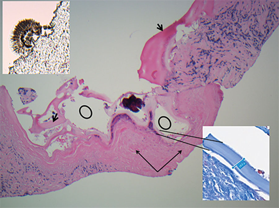
Figure 2. Kidney core biopsy of a child with disseminated Echinococcus multilocularis infection without liver involvement, Canada, 2018. Shown are folded laminated membrane (short black arrows) encircling variable-sized cystic structures (black circles) containing calcified and necrotic debris and dense periparasitic fibrosis (long black arrows), in a background of chronic inflammation and fibrosis. No residual normal kidney parenchyma was seen (hematoxylin and eosin stain, original magnification × 40). Inset at lower right shows laminar membrane, 18–19.4 μm in thickness in a background of fibrosis (Masson trichrome, original magnification ×40). Insert at upper left shows scolex attached to the paraffin edge of the block (original magnification ×40).
1All authors contributed equally to the preparation of this article.