Volume 27, Number 4—April 2021
Research
Histopathological Characterization of Cases of Spontaneous Fatal Feline Severe Fever with Thrombocytopenia Syndrome, Japan
Figure 4
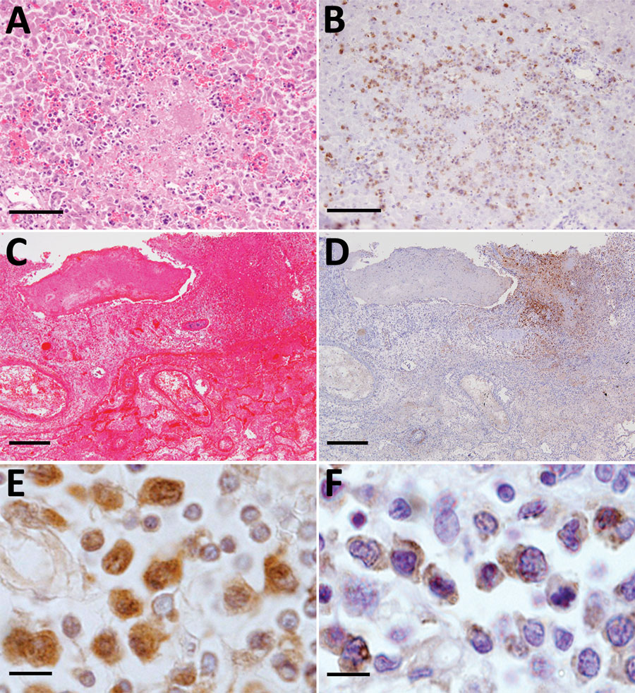
Figure 4. Necrotic foci in the liver and lung from fatal cases of severe fever with thrombocytopenia syndrome (SFTS) in cats, Japan. A, B) Hematoxylin & eosin (HE)–stained (A) and immunohistochemistry-stained (B) liver sections demonstrating SFTS virus–positive blastic lymphocytes in the necrotic foci. Scale bars indicate 100 μm. C, D) HE-stained (C) and immunohistochemistry-stained (D) lung sections demonstrating lymphocytes in the necrotic foci from the lungs. Scale bars indicates 200 μm. E, F) Ki67 (E) and Ig lambda chain (F) immunohistochemistry positively staining blastic lymphocytes. Scale bars indicates 10 μm.
1Current affiliation: National Institute of Infectious Diseases, Tokyo, Japan.
2Current affiliation: Okayama University of Science, Ehime, Japan.