Volume 27, Number 9—September 2021
Etymologia
Talaromyces marneffei
Figure 2
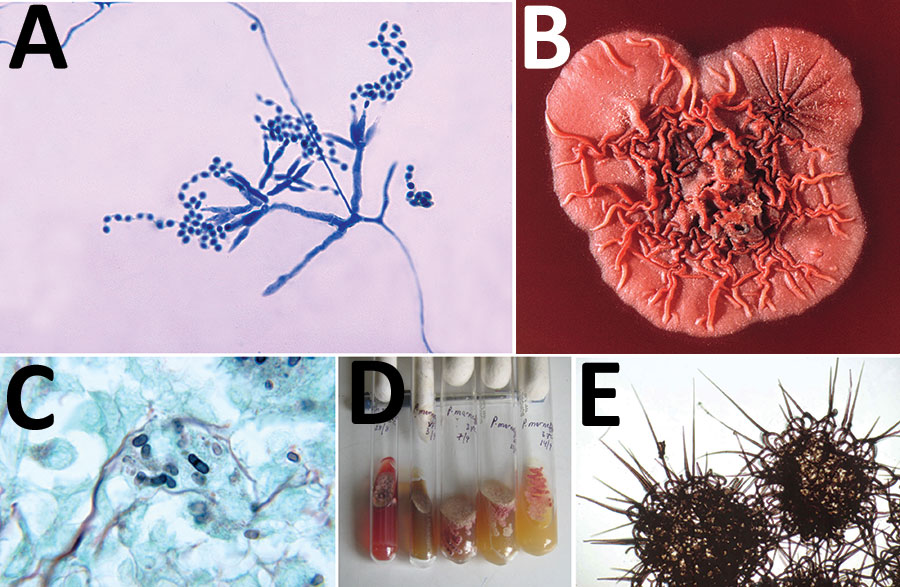
Figure 2. A) Ultrastructural morphology of Talaromyces marneffei, including chains of single-celled, teardrop-shaped conidia, each originating from its respective, flask-shaped phialide. Source: Libero Ajello, Centers for Disease Control and Prevention (https://phil.cdc.gov/Details.aspx?pid = 4240). B) Superior (front) view of a petri dish culture plate on which a wrinkled colony of Penicillium marneffei has been cultivated. Source: James Gathany, Centers for Disease Control and Prevention (https://phil.cdc.gov/Details.aspx?pid = 1879). C) Mouse testicle tissue specimen showing globe-shaped yeast cells of T. marneffei undergoing multiplication by binary fission not by mitosis (methenamine silver stain). Source: Libero Ajello, Centers for Disease Control and Prevention (https://phil.cdc.gov/Details.aspx?pid = 4235); D) Gradual conversion of mycelial phase of T. marneffei (growth at 25°C) to yeast phase on brain heart infusion agar after incubation at 37°C. Mycelial phase (first tube marked 25°C) shows diffusible red pigment. Source: Monika Mahajan, Postgraduate Institute of Medical Education and Research, Chandigarh, India; E) Loose network of hyphae of T. marneffei forming gymnothecium that contains asci. Source: https://istudy.pk/ascomycota-fruit-bodies/.