Volume 28, Number 4—April 2022
Dispatch
Rigidoporus corticola Colonization and Invasive Fungal Disease in Immunocompromised Patients, United States
Figure 1
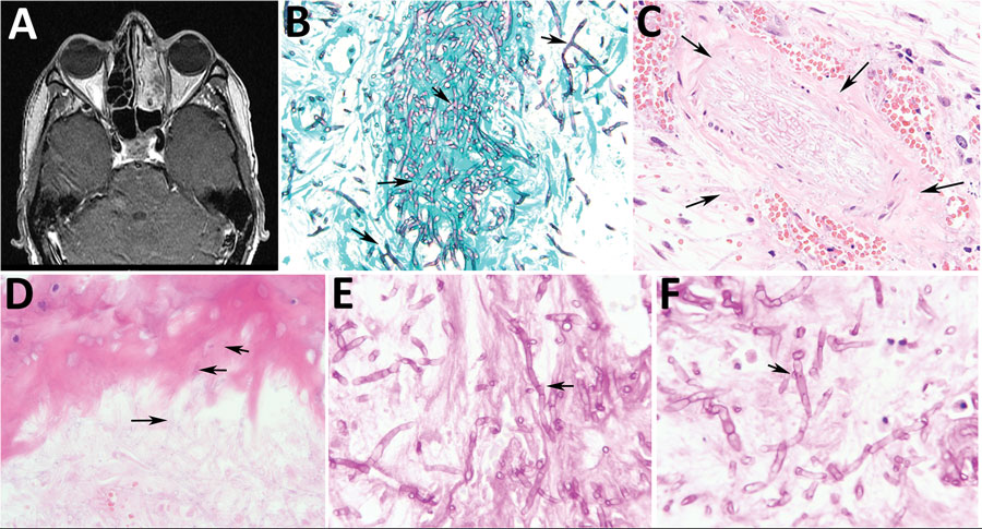
Figure 1. Radiologic and pathologic findings in a 43-year-old immunocompromised man with chronic granulomatous disease and diagnosed invasive fungal sinusitis caused by Rigidoporus corticola (Oxyporus corticola) infection, United States. A) Magnetic resonance imaging of the head showing mucosal thickening and near opacification in the left frontal, left ethmoid, left maxillary, and left sphenoid sinuses, and the left nasal cavity. No findings suggest extra-sinus extension. B) Debrided sinus tissue stained with Gomori methenamine silver. Arrows indicate numerous septate hyphae with predominantly right-angle branching. Original magnification ×600. C) Hematoxylin and eosin stain of blood vessels. Arrows indicate permeation (angioinvasion). Original magnification ×600. D) Hematoxylin and eosin stain of the sinonasal ossicles. Arrows indicate infiltration consistent with invasive fungal disease. Original magnification ×600. E, F) Periodic acid–Schiff-diastase–stained sinus tissue. Arrows indicate bulge-like (E) and hook-like (F) hyphal outgrowths potentially representing clamp connections, characteristic of filamentous basidiomycetes, that were detected upon careful examination. Original magnification ×1,000.