Volume 28, Number 4—April 2022
Research
Fatal Human Alphaherpesvirus 1 Infection in Free-Ranging Black-Tufted Marmosets in Anthropized Environments, Brazil, 2012–2019
Tais M. Wilson, Jana M. Ritter, Roosecelis B. Martines, Hannah A. Bullock, Pamela Fair, Kay W. Radford, Isabel L. Macêdo, Davi E.R. Sousa, Alexandra A.B. Gonçalves, Alessandro P. Romano, Pedro H.O. Passsos, Daniel G. Ramos, Gabriela R.T. Costa, Karina R.L.J. Cavalcante, Cristiano B. de Melo, Sherif R. Zaki, and Marcio B. Castro
Figure 5
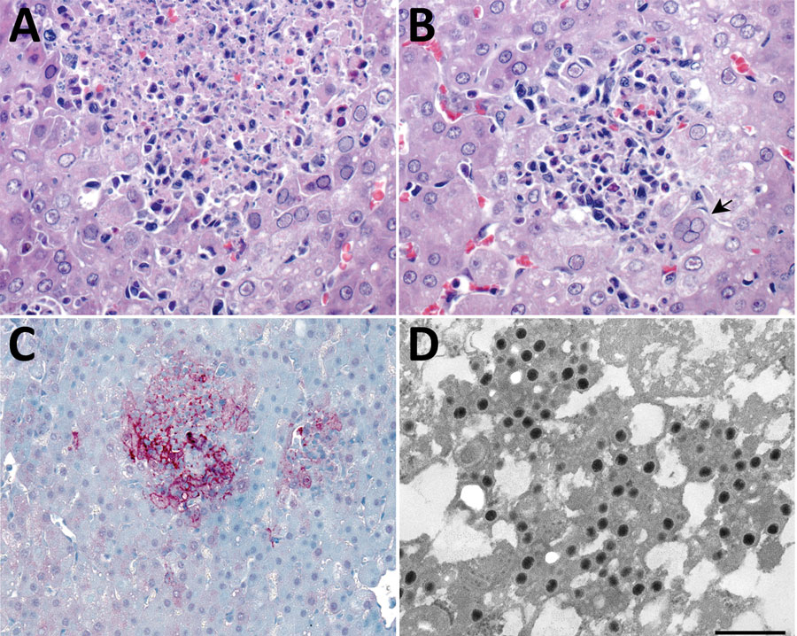
Figure 5. Liver pathology in free-ranging black-tufted marmosets with fatal human alphaherpesvirus 1 infection, Brazil, 2012–2019. A) Intranuclear inclusion bodies in hepatocytes at the margin of a necrotic focus. Hemotoxylin and eosin (H&E) stain; original magnification ×63. B) Multinucleated syncytial cell (arrow). H&E; original magnification ×63. C) Human alphaherpesvirus 1 immunostaining within hepatocytes in an area of necrotizing hepatitis. Immunihistochemistry; original magnification ×40. D) Herpesvirus in the cytoplasm of a hepatocyte. Transmission electron microscopy; scale bar indicates 400 nm.
Page created: January 26, 2022
Page updated: March 19, 2022
Page reviewed: March 19, 2022
The conclusions, findings, and opinions expressed by authors contributing to this journal do not necessarily reflect the official position of the U.S. Department of Health and Human Services, the Public Health Service, the Centers for Disease Control and Prevention, or the authors' affiliated institutions. Use of trade names is for identification only and does not imply endorsement by any of the groups named above.