Volume 30, Number 11—November 2024
Dispatch
Transmission of Severe Fever with Thrombocytopenia Syndrome Virus to Human from Nonindigenous Tick Host, Japan
Abstract
We report a human case of severe fever with thrombocytopenia syndrome virus infection transmitted by a tick, confirmed by viral identification. Haemaphysalis aborensis, a tick species not native to Japan that has been observed to transmit the virus to humans, is now recognized as a potential vector of this virus in Japan.
Blood-feeding ticks can transmit viruses to vertebrates, including humans. A previously unknown flavivirus, Saruyama virus, was detected in Japan in 2018 (1); similar viral sequences have also been identified in wild deer and boars in Japan. Severe fever with thrombocytopenia syndrome (SFTS) is an emerging tickborne disease caused by SFTS virus (SFTSV), which belongs to the family Phenuiviridae, genus Bandavirus. The first SFTS case was reported in China in 2010 (2), followed by cases in Japan and South Korea in 2013 (3,4); those 3 countries are the primary endemic areas for SFTSV.
Tick bites are the primary route of SFTSV transmission (2,5). Haemaphysalis longicornis ticks, native to east Asia, have been identified as a major SFTSV vector (2,6). SFTSV cases have been reported in several countries in Southeast Asia, including Vietnam and Thailand, and in South Asia, including Pakistan (7), suggesting that SFTSV might expand from endemic regions in tandem with its host animals or through tick migration. In Japan, several tick species, including H. flava, H. megaspinosa, H. kitaokai, H. formosensis, and H. hystricis, carry the SFTSV genome (8,9). The mortality rate for SFTS infection ranges from 5% to 28%; the elderly are at higher risk for fatal clinical outcomes (10), indicating its potential public health consequences. No antiviral drugs or vaccines are available for SFTSV infection.
We report a human case of SFTS transmitted by a novel tick host, H. aborensis, a tick not endemic to Japan that has been identified as a potential vector of SFTSV, indicating possible expansion of habitats of infectious ticks. That finding highlights the importance of comprehensive viral genome analysis as part of routine tick-borne viral surveillance. We obtained informed consent for publication from the patient and ethics approval from the Clinical Research Ethics Committee of Nagasaki University Hospital (record no. 23112012).
An 80-year-old female patient with a medical history of hypertension, bronchial asthma, and cerebral aneurysm (postoperative), but no history of smoking, alcohol consumption, or recent travel, experienced fever and dizziness. The case-patient resided in Nagasaki, Japan, close to the forest, and reported that she frequently encountered wild animals, such as wild boars and civets. She engaged in daily activities, regularly tended her garden, and had no companion animals. She sought medical consultation with a primary care physician on day 3 after onset of symptoms.
Blood tests revealed a drop in her leukocyte count to 3,180 cells/μL (reference range 3,300–8,600 cells/μL) and platelet count to 104,000/μL (reference range 158,000–348,000/μL). Subsequent blood tests showed further decreases in leukocytes to 1,690 cells/μL and platelets to 72,000/μL. On day 5, the case-patient was referred to a secondary emergency hospital for further evaluation. A blood-engorged tick was found on her inner right thigh (Figure 1). Presence of leukopenia, thrombocytopenia, and tick bites indicated SFTSV infection. A serum specimen from the patient was sent to the laboratory at Nagasaki City Health Center, which is responsible for administrative inspections for SFTS diagnosis. The results revealed SFTSV positivity on day 12 after symptom onset.
The patient was transferred from the secondary hospital to the Department of Internal Medicine of Infectious Diseases at Nagasaki University Hospital, a tertiary emergency hospital, on day 8 after onset. Physical examination at time of admission indicated a body temperature of 36.0°C, heartbeat of 66 beats/min, blood pressure of 121/72 mm Hg, SpO2 of 94% (room air), and respiratory rate of 28 breaths/min. The patient’s level of consciousness was unclear, but she responded when called, which is indicative of a II-10 rating on the Japan Coma Scale. Blood chemical examination demonstrated results with reference ranges for hemoglobin (12.3 g/dL), sodium (137 mEq/L), potassium (3.8 mEq/L), chloride (108 mEq/L), blood urea nitrogen (6 mg/dL), creatinine (0.7 mg/dL), and C-reactive protein (0.04 mg/dL). Compared with earlier test results, we noted further decreases in leukocyte count, to 2,300 cells/μL (neutrophils 920 cells/μL, lymphocytes 1,080 cells/μL), and platelet count, to 53,000/μL; we also saw increases in aspartate transferase (133 U/L, reference range 13–30 U/L), alanine transaminase (64 U/L, reference range 7–23 U/L), lactate dehydrogenase (770U/L, reference range 124–222 U/L), and creatine kinase (471 U/L, reference range 41–153 U/L). Urine examination revealed high protein 2+ and occult blood 2+ results. Results of blood cultures on days 6 and 10 and urine cultures on day 10 after onset were negative for bacterial infections. The patient gradually recovered and was discharged on day 29 after symptom onset without any specified lasting effects.
We sent the tick from the patient and serum specimens collected on days 6, 9, 10, and 17 after onset to the Department of Virology, Institute of Tropical Medicine, at Nagasaki University for examination. We extracted total RNA from the homogenized tick sample (Appendix) and subjected serum specimens to quantitative reverse-transcription PCR (qRT-PCR) (Appendix). The specimen from day 6 demonstrated the highest number of SFTSV RNA copies (1.06 × 10⁵/5 μL). The SFTSV RNA copies in the serum specimens decreased and were undetectable on day 17 after onset (Figure 1). Homogenates from the tick demonstrated a substantially higher number of SFTSV RNA copies (1.01 × 10⁷/5 μL) than the patient samples. We isolated viruses only from the tick, not from patient specimens.
To explore the genomic similarity of SFTSV strains derived from tick and human samples, we determined the full-length protein-coding sequences of the large (L), medium (M), and small (S) segments of viruses from the tick (GenBank accession nos. PP813867–9) and patient (accession nos. PP839300–2) by using next-generation sequencing (Appendix). We conducted phylogenetic analysis using MEGA11 (https://www.megasoftware.net) to determine the genetic relationships between the sequences from our study and previously identified SFTSV sequences from countries in Asia, including Japan (11). The sequences of patient- and tick-derived SFTS L/M/S segments were identical. SFTSV identified in our study’s belonged to B-2 clade (Figure 2, panels A–C), the genotype most prevalent in Japan and South Korea (10).
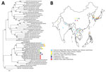
Morphologic characteristics (Figure 1) identified the tick collected from the patient as belonging to the genus Haemaphysalis. To confirm species identification, we sequenced the 16S ribosomal RNA (accession no. PP813416) (Appendix). Phylogenetic analysis identified it as most closely related to H. aborensis, a species not endemic to Japan (Figure 3, panel A). We found no previous reports of SFTSV isolation or gene detection in H. aborensis ticks.
H. aborensis ticks are primarily distributed in Nepal and India in South Asia and Laos, Vietnam, and Thailand in Southeast Asia (Figure 3, panel B) (12); porcupines, wild boars, and deer are the primary hosts (12). A previous study identified H. aborensis ticks collected from Turdus pallidus (pale thrush) on Hong Island, South Korea (13). The T. pallidus thrush is a migratory bird that breeds in areas from northeast China to far eastern Russia and overwinters in southern and central Japan, South Korea, and southern China (14). Because the B-2 clade has been isolated only in Japan and South Korea (10), SFTSV-infected ticks were likely carried by infected birds from South Korea. Although it is possible that ticks were carried by birds from South Korea, then acquired and transmitted SFTSV through infected animals in Japan, this scenario is unlikely because H. aborensis ticks had not been previously identified in Japan.
We report a case of tick-transmitted SFTSV infection in a human patient. Virus isolation and identification of the tick species confirmed that H. aborensis ticks can transmit SFTSV to humans. The phylogenetic analysis revealed no differences between sequences of SFTSV from the tick and the patient. Identifying an additional host tick highlights the importance of routine tick surveillance for monitoring SFTSV expansion.
Mr. Xu is a doctoral student in the Program for Nurturing Global Leaders in Tropical and Emerging Communicable Diseases at the Graduate School of Biomedical Sciences, Nagasaki University, Nagasaki, Japan. His research interests include the epidemiology of SFTSV and the elucidation of the molecular mechanism of the SFTSV replication cycle.
Acknowledgments
The authors acknowledge Katsuaki Motomiya for contributing to obtaining patient specimens and Mitsuru Hattori and Akira Yoshikawa for fruitful discussions. The authors acknowledge Tomomi Kurashige and Megumi Tsubota for their technical support and all members of the Department of Virology, Institute of Tropical Medicine, Nagasaki University for their cooperation.
This study was supported by the Japan Agency of Medical Research and Development (grant nos. JP24fk0108656, JP24fk0108695, JP24wm0125006, JP24wm0125011, JP23fm0208101, JP23fk0108656, JP23wm0125006, JP22wm0325023, JP22fm0208101, JP21fm0208101, JP21wm0325023, and JP20wm0323023); the Japan Society for Promotion of Sciences (grant nos. 21K07059, 22KK0115, and 24K02288); Takeda Science Foundation, MSD Life Science Foundation, the Naito Foundation, Kurozumi Medical Foundation, the Asahi Glass Foundation, and Joint/Research Center on Tropical Disease, Institute of Tropical Medicine, Nagasaki University (2022-Ippan-12, 2023-Ippan-16).
References
- Kobayashi D, Inoue Y, Suzuki R, Matsuda M, Shimoda H, Faizah AN, et al. Identification and epidemiological study of an uncultured flavivirus from ticks using viral metagenomics and pseudoinfectious viral particles. Proc Natl Acad Sci U S A. 2024;121:
e2319400121 . DOIPubMedGoogle Scholar - Yu XJ, Liang MF, Zhang SY, Liu Y, Li JD, Sun YL, et al. Fever with thrombocytopenia associated with a novel bunyavirus in China. N Engl J Med. 2011;364:1523–32. DOIPubMedGoogle Scholar
- Takahashi T, Maeda K, Suzuki T, Ishido A, Shigeoka T, Tominaga T, et al. The first identification and retrospective study of Severe Fever with Thrombocytopenia Syndrome in Japan. J Infect Dis. 2014;209:816–27. DOIPubMedGoogle Scholar
- Kim YR, Yun Y, Bae SG, Park D, Kim S, Lee JM, et al. Severe fever with thrombocytopenia syndrome virus infection, South Korea, 2010. Emerg Infect Dis. 2018;24:2103–5. DOIPubMedGoogle Scholar
- Yun SM, Lee WG, Ryou J, Yang SC, Park SW, Roh JY, et al. Severe fever with thrombocytopenia syndrome virus in ticks collected from humans, South Korea, 2013. Emerg Infect Dis. 2014;20:1358–61. DOIPubMedGoogle Scholar
- Park SW, Song BG, Shin EH, Yun SM, Han MG, Park MY, et al. Prevalence of severe fever with thrombocytopenia syndrome virus in Haemaphysalis longicornis ticks in South Korea. Ticks Tick Borne Dis. 2014;5:975–7. DOIPubMedGoogle Scholar
- Kim EH, Park SJ. Emerging tick-borne Dabie bandavirus: virology, epidemiology, and prevention. Microorganisms. 2023;11:2309. DOIPubMedGoogle Scholar
- Sato Y, Mekata H, Sudaryatma PE, Kirino Y, Yamamoto S, Ando S, et al. Isolation of severe fever with thrombocytopenia syndrome virus from various tick species in area with human severe fever with thrombocytopenia syndrome cases. Vector Borne Zoonotic Dis. 2021;21:378–84. DOIPubMedGoogle Scholar
- National Institute of Infectious Diseases. Japan. Infectious agents surveillance report, volume 37. Severe fever with thrombocytopenia syndrome (SFTS) in Japan, as of February 2016 [cited 2024 Jun 5]. https://www.niid.go.jp/niid/en/iasr-vol37-e/865-iasr/6339-tpc433.html
- Casel MA, Park SJ, Choi YK. Severe fever with thrombocytopenia syndrome virus: emerging novel phlebovirus and their control strategy. Exp Mol Med. 2021;53:713–22. DOIPubMedGoogle Scholar
- Yun SM, Park SJ, Kim YI, Park SW, Yu MA, Kwon HI, et al. Genetic and pathogenic diversity of severe fever with thrombocytopenia syndrome virus (SFTSV) in South Korea. JCI Insight. 2020;5:
e129531 . DOIPubMedGoogle Scholar - Hoogstraal H, Dhanda V, Kammah KME. Aborphysalis, a new subgenus of Asian Haemaphysalis ticks; and identity, distribution, and hosts of H. aborensis Warburton (resurrected ) (Ixodoidea: Ixodidae). J Parasitol. 1971;57:748–60. DOIPubMedGoogle Scholar
- Kim HC, Chong ST, Choi CY, Nam HY, Chae HY, Klein TA, et al. Tick surveillance, including new records for three Haemaphysalis species (Acari: Ixodidae) collected from migratory birds during 2009 on Hong Island (Hong-do), Republic of Korea. Syst Appl Acarol. 2016;21:596–606. DOIGoogle Scholar
- Collar N, de Juana E. Birds of the world 2020. Pale thrush (Turdus pallidus) [cited 2024 Jun 6]. https://birdsoftheworld.org/bow/species/palthr1/cur/introduction
Figures
Cite This ArticleOriginal Publication Date: October 20, 2024
Table of Contents – Volume 30, Number 11—November 2024
| EID Search Options |
|---|
|
|
|
|
|
|


Please use the form below to submit correspondence to the authors or contact them at the following address:
Yuki Takamatsu, Department of Virology, Institute of Tropical Medicine, Nagasaki University, 1-12-4 Sakamoto, Nagasaki 852-8523, Japan
Top