Disclaimer: Early release articles are not considered as final versions. Any changes will be reflected in the online version in the month the article is officially released.
Volume 31, Number 5—May 2025
Dispatch
Administration of L-type Bovine Spongiform Encephalopathy to Macaques to Evaluate Zoonotic Potential
Figure 2
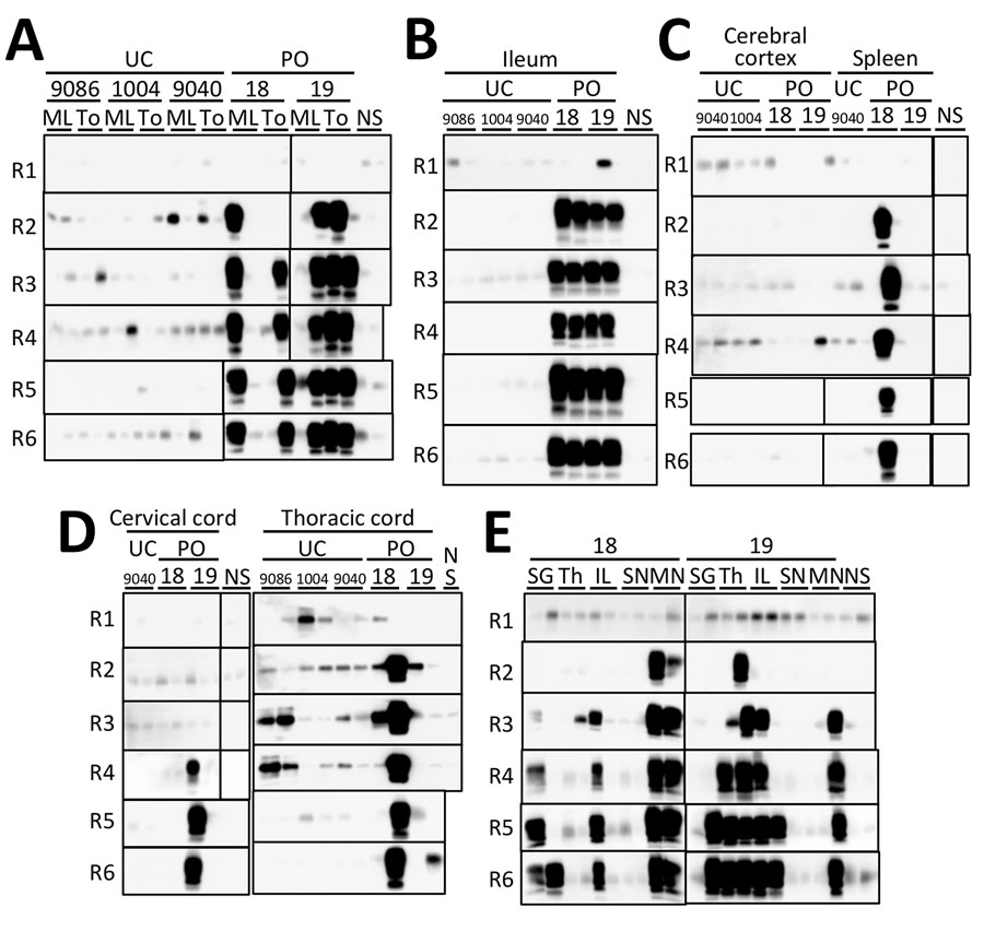
Figure 2. Sensitivity of modified protein misfolding cyclic amplification (PMCA) to detect abnormal prion protein in study of oral transmission of L-type bovine spongiform encephalopathy (L-BSE) in orally inoculated macaques to evaluate zoonotic potential. We performed 6 rounds of PMCA in duplicate in the tissues of 2 macaques (#18 and #19) orally inoculated with L-BSE prions, primarily in the lymphoid and nervous system tissues. A) ML nodes and To tissue; B) ileum; C) cerebral cortex and spleen; D) cervical and thoracic cords; and E) SG, Th, IL nodes, SN, and MN tissues. We prepared PMCA seeds, equivalent to 6.25 mg of tissue, obtained from lymphoid tissues, peripheral nerves, submaxillary glands, and ileum by using a standard sodium phosphotungstic acid precipitation method. For brain tissues, we used 10% homogenates in phosphate-buffered saline (250 μg) and phosphotungstic acid precipitates as seeds. For spinal cords, we used 10% homogenates in phosphate-buffered saline and ethanol precipitates (625 μg equivalent) as seeds. Tissues obtained from 3 uninfected macaques (#9068, #1004, and #9040) served as negative controls and were processed identically to those obtained from inoculated animals. For the thoracic spinal cord of macaque #19, we performed amplification for 7 rounds to determine whether the round 6 signal was positive. IL, inguinal lymph; ML, mesenteric lymph; MN, median nerve; NS, nonseeded control; PMCA, protein misfolding cyclic amplification; PO, L-type bovine spongiform encephalopathy orally inoculated macaques; SG, submaxillary gland; SN, sciatic nerve; TH, thymus; To, tonsil; UC, uninoculated control.
1These authors were co–principal investigators.