Disclaimer: Early release articles are not considered as final versions. Any changes will be reflected in the online version in the month the article is officially released.
Volume 31, Number 5—May 2025
Research Letter
Trichophyton indotineae Infection, São Paulo, Brazil, 2024
João Nobrega de Almeida , Amanda R. dos Santos, Mario Roberto de S. Trindade, Jeremy A. W. Gold, Fernanda P.M. Razo, Sarah S. Gonçalves, Erick G. Dorlass, Renato de Mello Ruiz, Jacyr Pasternak, Cristovão L.P. Mangueira, Silke Uhrlaß, Pietro Nenoff, Shyam B. Verma, André M. Doi, and Marines D.V. Martino
Figure 1
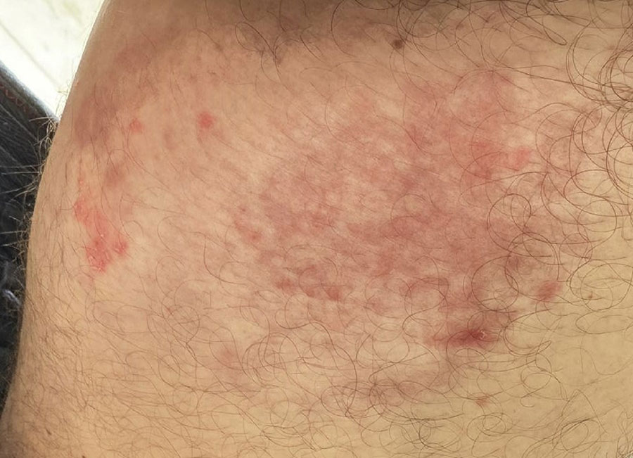
Figure 1. Atypical tinea cruris from Trichophyton indotineae infection, São Paulo, Brazil, 2024. A photograph of the left groin (provided by the patient) shows lesions characterized by poorly defined margins, hyperemic scaly plaques in the medial region, and an inflammatory infiltrate in the central-lateral area.
Page created: April 03, 2025
Page updated: April 22, 2025
Page reviewed: April 22, 2025
The conclusions, findings, and opinions expressed by authors contributing to this journal do not necessarily reflect the official position of the U.S. Department of Health and Human Services, the Public Health Service, the Centers for Disease Control and Prevention, or the authors' affiliated institutions. Use of trade names is for identification only and does not imply endorsement by any of the groups named above.