Perspective
International Editor's Update
| EID | Kurata T. International Editor's Update. Emerg Infect Dis. 2000;6(6):565. https://doi.org/10.3201/eid0606.000601 |
|---|---|
| AMA | Kurata T. International Editor's Update. Emerging Infectious Diseases. 2000;6(6):565. doi:10.3201/eid0606.000601. |
| APA | Kurata, T. (2000). International Editor's Update. Emerging Infectious Diseases, 6(6), 565. https://doi.org/10.3201/eid0606.000601. |
Recent Trends in Tuberculosis, Japan
Despite a decline after World War II, the rate of tuberculosis in Japan remains high. Infection is heavily concentrated in the >60-year age group, and 82% of patients are >40 years of age. The success rate for treatment of smear-positive patients is 78%. Multidrug-resistant strains of Mycobacterium tuberculosis are rare.
| EID | Mori T. Recent Trends in Tuberculosis, Japan. Emerg Infect Dis. 2000;6(6):566-568. https://doi.org/10.3201/eid0606.000602 |
|---|---|
| AMA | Mori T. Recent Trends in Tuberculosis, Japan. Emerging Infectious Diseases. 2000;6(6):566-568. doi:10.3201/eid0606.000602. |
| APA | Mori, T. (2000). Recent Trends in Tuberculosis, Japan. Emerging Infectious Diseases, 6(6), 566-568. https://doi.org/10.3201/eid0606.000602. |
Trends in Flavivirus Infections in Japan
Although Japanese encephalitis has declined as an important cause of illness and death in Japan, infection with other flaviviruses has become a public health concern. Recently, reports of imported dengue cases, as well as isolations of tick-borne encephalitis virus, have increased.
| EID | Kurane I, Takasaki T, Yamada K. Trends in Flavivirus Infections in Japan. Emerg Infect Dis. 2000;6(6):569-571. https://doi.org/10.3201/eid0606.000603 |
|---|---|
| AMA | Kurane I, Takasaki T, Yamada K. Trends in Flavivirus Infections in Japan. Emerging Infectious Diseases. 2000;6(6):569-571. doi:10.3201/eid0606.000603. |
| APA | Kurane, I., Takasaki, T., & Yamada, K. (2000). Trends in Flavivirus Infections in Japan. Emerging Infectious Diseases, 6(6), 569-571. https://doi.org/10.3201/eid0606.000603. |
Trends in Antimicrobial-Drug Resistance in Japan
Multidrug resistance in gram-positive bacteria has become common worldwide. In Japan until recently, gram-negative bacteria such as Pseudomonas aeruginosa, Klebsiella pneumoniae, and Serratia marcescens were controlled by carbapenems, fluoroquinolones, and aminoglycosides. However, several of these microorganisms have recently developed resistance against many antimicrobial drugs.
| EID | Arakawa Y, Ike Y, Nagasawa M, Shibata N, Doi Y, Shibayama K, et al. Trends in Antimicrobial-Drug Resistance in Japan. Emerg Infect Dis. 2000;6(6):572-575. https://doi.org/10.3201/eid0606.000604 |
|---|---|
| AMA | Arakawa Y, Ike Y, Nagasawa M, et al. Trends in Antimicrobial-Drug Resistance in Japan. Emerging Infectious Diseases. 2000;6(6):572-575. doi:10.3201/eid0606.000604. |
| APA | Arakawa, Y., Ike, Y., Nagasawa, M., Shibata, N., Doi, Y., Shibayama, K....Kurata, T. (2000). Trends in Antimicrobial-Drug Resistance in Japan. Emerging Infectious Diseases, 6(6), 572-575. https://doi.org/10.3201/eid0606.000604. |
Developing National Epidemiological Capacity to Meet the Challenges of Emerging Infections in Germany
In January 1996, the Robert Koch Institute, Germany’s national public health institute, began strengthening its epidemiologic capacity to respond to emerging and other infectious diseases. Six integrated strategies were initiated: developing employee training, outbreak investigation, and epidemiologic research programs; strengthening surveillance systems; improving communications to program partners and constituents; and building international collaborations. By December 1999, five employees had completed a 2-year applied epidemiology training program, 186 health department personnel had completed a 2-week training course, 27 outbreak investigations had been completed, eight short-term research projects had been initiated, major surveillance and epidemiologic research efforts for foodborne and nosocomial infections had begun, and 16 scientific manuscripts had been published or were in press. The German experience indicates that, with a concerted effort, considerable progress in building a national applied infectious disease program can be achieved in a short time frame.
| EID | Petersen LR, Ammon A, Hamouda O, Breuer T, Kießling S, Bellach B, et al. Developing National Epidemiological Capacity to Meet the Challenges of Emerging Infections in Germany. Emerg Infect Dis. 2000;6(6):576-584. https://doi.org/10.3201/eid0606.000605 |
|---|---|
| AMA | Petersen LR, Ammon A, Hamouda O, et al. Developing National Epidemiological Capacity to Meet the Challenges of Emerging Infections in Germany. Emerging Infectious Diseases. 2000;6(6):576-584. doi:10.3201/eid0606.000605. |
| APA | Petersen, L. R., Ammon, A., Hamouda, O., Breuer, T., Kießling, S., Bellach, B....Kurth, R. (2000). Developing National Epidemiological Capacity to Meet the Challenges of Emerging Infections in Germany. Emerging Infectious Diseases, 6(6), 576-584. https://doi.org/10.3201/eid0606.000605. |
Evidence Against Rapid Emergence of Praziquantel Resistance in Schistosoma haematobium, Kenya
We examined the long-term efficacy of praziquantel against Schistosoma haematobium, the causative agent of urinary schistosomiasis, during a school-based treatment program in the Msambweni area of Coast Province, Kenya, where the disease is highly endemic. Our results, derived from treating 4,031 of 7,641 children from 1984 to 1993, indicate substantial year-to-year variation in drug efficacy. However, the pattern of this variation was not consistent with primary or progressive emergence of praziquantel resistance. Mathematical modeling indicated that, at current treatment rates, praziquantel resistance will likely take 10 or more years to emerge.
| EID | King CH, Muchiri EM, Ouma JH. Evidence Against Rapid Emergence of Praziquantel Resistance in Schistosoma haematobium, Kenya. Emerg Infect Dis. 2000;6(6):585-594. https://doi.org/10.3201/eid0606.000606 |
|---|---|
| AMA | King CH, Muchiri EM, Ouma JH. Evidence Against Rapid Emergence of Praziquantel Resistance in Schistosoma haematobium, Kenya. Emerging Infectious Diseases. 2000;6(6):585-594. doi:10.3201/eid0606.000606. |
| APA | King, C. H., Muchiri, E. M., & Ouma, J. H. (2000). Evidence Against Rapid Emergence of Praziquantel Resistance in Schistosoma haematobium, Kenya. Emerging Infectious Diseases, 6(6), 585-594. https://doi.org/10.3201/eid0606.000606. |
Investigating Disease Outbreaks under a Protocol to the Biological and Toxin Weapons Convention
The Biological and Toxin Weapons Convention prohibits the development, production, and stockpiling of biological weapons agents or delivery devices for anything other than peaceful purposes. A protocol currently in the final stages of negotiation adds verification measures to the convention. One of these measures will be international investigation of disease outbreaks that suggest a violation of the convention, i.e., outbreaks that may be caused by use of biological weapons or release of harmful agents from a facility conducting prohibited work. Adding verification measures to the current Biological and Toxin Weapons Convention will affect the international public health and epidemiology communities; therefore, active involvement of these communities in planning the implementation details of the protocol will be important.
| EID | Wheelis M. Investigating Disease Outbreaks under a Protocol to the Biological and Toxin Weapons Convention. Emerg Infect Dis. 2000;6(6):595-600. https://doi.org/10.3201/eid0606.000607 |
|---|---|
| AMA | Wheelis M. Investigating Disease Outbreaks under a Protocol to the Biological and Toxin Weapons Convention. Emerging Infectious Diseases. 2000;6(6):595-600. doi:10.3201/eid0606.000607. |
| APA | Wheelis, M. (2000). Investigating Disease Outbreaks under a Protocol to the Biological and Toxin Weapons Convention. Emerging Infectious Diseases, 6(6), 595-600. https://doi.org/10.3201/eid0606.000607. |
Synopses
Hemophagocytic Syndromes and Infection
Hemophagocytic lymphohistiocytosis (HLH) is an unusual syndrome characterized by fever, splenomegaly, jaundice, and the pathologic finding of hemophagocytosis (phagocytosis by macrophages of erythrocytes, leukocytes, platelets, and their precursors) in bone marrow and other tissues. HLH may be diagnosed in association with malignant, genetic, or autoimmune diseases but is also prominently linked with Epstein-Barr (EBV) virus infection. Hyperproduction of cytokines, including interferon-γ and tumor necrosis factor-α, by EBV-infected T lymphocytes may play a role in the pathogenesis of HLH. EBV-associated HLH may mimic T-cell lymphoma and is treated with cytotoxic chemotherapy, while hemophagocytic syndromes associated with nonviral pathogens often respond to treatment of the underlying infection.
| EID | Fisman DN. Hemophagocytic Syndromes and Infection. Emerg Infect Dis. 2000;6(6):601-608. https://doi.org/10.3201/eid0606.000608 |
|---|---|
| AMA | Fisman DN. Hemophagocytic Syndromes and Infection. Emerging Infectious Diseases. 2000;6(6):601-608. doi:10.3201/eid0606.000608. |
| APA | Fisman, D. N. (2000). Hemophagocytic Syndromes and Infection. Emerging Infectious Diseases, 6(6), 601-608. https://doi.org/10.3201/eid0606.000608. |
Research
Predominance of HIV-1 Subtype A and D Infections in Uganda
To better characterize the virus isolates associated with the HIV-1 epidemic in Uganda, 100 specimens from HIV-1-infected persons were randomly selected from each of two periods from late 1994 to late 1997. The 200 specimens were classified into HIV-1 subtypes by sequence-based phylogenetic analysis of the envelope (env) gp41 region; 98 (49%) were classified as env subtype A, 96 (48%) as D, 5 (2.5%) as C, and 1 was not classified as a known env subtype. Demographic characteristics of persons infected with the two principal HIV-1 subtypes, A and D, were very similar, and the proportion of either subtype did not differ significantly between early and later periods. Our systematic characterization of the HIV-1 epidemic in Uganda over an almost 3-year period documented that the distribution and degree of genetic diversity of the HIV subtypes A and D are very similar and have not changed appreciably over that time.
| EID | Hu DJ, Baggs J, Downing RG, Pieniazek D, Dorn J, Fridlund C, et al. Predominance of HIV-1 Subtype A and D Infections in Uganda. Emerg Infect Dis. 2000;6(6):509-515. https://doi.org/10.3201/eid0606.000609 |
|---|---|
| AMA | Hu DJ, Baggs J, Downing RG, et al. Predominance of HIV-1 Subtype A and D Infections in Uganda. Emerging Infectious Diseases. 2000;6(6):509-515. doi:10.3201/eid0606.000609. |
| APA | Hu, D. J., Baggs, J., Downing, R. G., Pieniazek, D., Dorn, J., Fridlund, C....Lal, R. B. (2000). Predominance of HIV-1 Subtype A and D Infections in Uganda. Emerging Infectious Diseases, 6(6), 509-515. https://doi.org/10.3201/eid0606.000609. |
Hantavirus Pulmonary Syndrome Associated with Monongahela Virus, Pennsylvania
The first two recognized cases of rapidly fatal hantavirus pulmonary syndrome in Pennsylvania occurred within an 8-month period in 1997. Illness in the two patients was confirmed by immunohistochemical techniques on autopsy material. Reverse transcription-polymerase chain reaction analysis of tissue from one patient and environmentally associated Peromyscus leucopus (white-footed mouse) identified the Monongahela virus variant. Physicians should be vigilant for such Monongahela virus-associated cases in the eastern United States and Canada, particularly in the Appalachian region.
| EID | Rhodes LV, Huang C, Sanchez AJ, Nichol ST, Zaki SR, Ksiazek TG, et al. Hantavirus Pulmonary Syndrome Associated with Monongahela Virus, Pennsylvania. Emerg Infect Dis. 2000;6(6):616-621. https://doi.org/10.3201/eid0606.000610 |
|---|---|
| AMA | Rhodes LV, Huang C, Sanchez AJ, et al. Hantavirus Pulmonary Syndrome Associated with Monongahela Virus, Pennsylvania. Emerging Infectious Diseases. 2000;6(6):616-621. doi:10.3201/eid0606.000610. |
| APA | Rhodes, L. V., Huang, C., Sanchez, A. J., Nichol, S. T., Zaki, S. R., Ksiazek, T. G....Knecht, K. R. (2000). Hantavirus Pulmonary Syndrome Associated with Monongahela Virus, Pennsylvania. Emerging Infectious Diseases, 6(6), 616-621. https://doi.org/10.3201/eid0606.000610. |
Risk Factors for Otitis Media and Carriage of Multiple Strains of Haemophilus influenzae and Streptococcus pneumoniae
We studied genetic diversity in Streptococcus pneumoniae and Haemophilus influenzae in throat culture isolates from 38 children attending two day-care centers in Michigan. Culture specimens were collected weekly; 184 S. pneumoniae and 418 H. influenzae were isolated from the cultures. Pulsed-field gel electrophoresis identified 29 patterns among the S. pneumoniae isolates and 87 among the H. influenzae isolates. Of the cultures, 5% contained multiple genetic types of S. pneumoniae, and 43% contained multiple types of H. influenzae. Carriage of multiple H. influenzae isolates, which was associated with exposure to smoking, history of allergies, and age 36 to 47 months, may increase risk for otitis media in children.
| EID | St. Sauver J, Marrs CF, Foxman B, Somsel P, Madera R, Gilsdorf JR. Risk Factors for Otitis Media and Carriage of Multiple Strains of Haemophilus influenzae and Streptococcus pneumoniae. Emerg Infect Dis. 2000;6(6):622-630. https://doi.org/10.3201/eid0606.000611 |
|---|---|
| AMA | St. Sauver J, Marrs CF, Foxman B, et al. Risk Factors for Otitis Media and Carriage of Multiple Strains of Haemophilus influenzae and Streptococcus pneumoniae. Emerging Infectious Diseases. 2000;6(6):622-630. doi:10.3201/eid0606.000611. |
| APA | St. Sauver, J., Marrs, C. F., Foxman, B., Somsel, P., Madera, R., & Gilsdorf, J. R. (2000). Risk Factors for Otitis Media and Carriage of Multiple Strains of Haemophilus influenzae and Streptococcus pneumoniae. Emerging Infectious Diseases, 6(6), 622-630. https://doi.org/10.3201/eid0606.000611. |
Molecular Evidence of Clonal Vibrio parahaemolyticus Pandemic Strains
The upsurge in worldwide incidence of Vibrio parahaemolyticus infection in the last 5 years has been attributed to the recent appearance of three serotypes with pandemic potential: O3:K6, O4:K68, and O1:K untypeable (KUT). Thirty-five strains of these serotypes, isolated from different countries over 4 years, were characterized by ribotyping and pulsed-field gel electrophoresis to determine their origin. The ribotypes of the strains of these serotypes were indistinguishable, except for a Japanese tdh- negative O3:K6 strain and a U.S. clinical O3:K6 isolate, which had slightly different profiles. The migration patterns of the NotI-digest of the total DNA of the strains were similar, and only slight variations were observed between the serotypes. By contrast, the O3:K6 and O1:KUT strains isolated before 1995 and strains of other serotypes had markedly different profiles. The O4:K68 and O1:KUT strains most likely originated from the pandemic O3:K6 clone.
| EID | Chowdhury NR, Chakraborty S, Ramamurthy T, Nishibuchi M, Yamasaki S, Takeda Y, et al. Molecular Evidence of Clonal Vibrio parahaemolyticus Pandemic Strains. Emerg Infect Dis. 2000;6(6):631-636. https://doi.org/10.3201/eid0606.000612 |
|---|---|
| AMA | Chowdhury NR, Chakraborty S, Ramamurthy T, et al. Molecular Evidence of Clonal Vibrio parahaemolyticus Pandemic Strains. Emerging Infectious Diseases. 2000;6(6):631-636. doi:10.3201/eid0606.000612. |
| APA | Chowdhury, N. R., Chakraborty, S., Ramamurthy, T., Nishibuchi, M., Yamasaki, S., Takeda, Y....Nair, G. B. (2000). Molecular Evidence of Clonal Vibrio parahaemolyticus Pandemic Strains. Emerging Infectious Diseases, 6(6), 631-636. https://doi.org/10.3201/eid0606.000612. |
Dispatches
Mass Die-Off of Caspian Seals Caused by Canine Distemper Virus
Thousands of Caspian seals (Phoca caspica) died in the Caspian Sea from April to August 2000. Lesions characteristic of morbillivirus infection were found in tissue specimens from dead seals. Canine distemper virus infection was identified by serologic examination, reverse transcriptase-polymerase chain reaction, and sequencing of selected P gene fragments. These results implicate canine distemper virus infection as the primary cause of death.
| EID | Kennedy S, Kuiken T, Jepson PD, Deaville R, Forsyth M, Barrett T, et al. Mass Die-Off of Caspian Seals Caused by Canine Distemper Virus. Emerg Infect Dis. 2000;6(6):637-639. https://doi.org/10.3201/eid0606.000613 |
|---|---|
| AMA | Kennedy S, Kuiken T, Jepson PD, et al. Mass Die-Off of Caspian Seals Caused by Canine Distemper Virus. Emerging Infectious Diseases. 2000;6(6):637-639. doi:10.3201/eid0606.000613. |
| APA | Kennedy, S., Kuiken, T., Jepson, P. D., Deaville, R., Forsyth, M., Barrett, T....Wilson, S. (2000). Mass Die-Off of Caspian Seals Caused by Canine Distemper Virus. Emerging Infectious Diseases, 6(6), 637-639. https://doi.org/10.3201/eid0606.000613. |
Nontoxigenic Corynebacterium diphtheriae: An Emerging Pathogen in England and Wales?
Confirmed isolates of nontoxigenic Corynebacterium diphtheriae in England and Wales increased substantially from 1986 to 1994. Ribotyping of 121 isolates confirmed in 1995 showed that 90 were of a single strain isolated exclusively from the throat; none had previously been identified in toxigenic strains from U.K. or non-U.K. residents. The upward trend in nontoxigenic C. diphtheriae probably represented increased ascertainment, although dissemination of a particular strain or clone may have been a factor.
| EID | Reacher M, Ramsay M, White J, De Zoysa A, Efstratiou A, Mann G, et al. Nontoxigenic Corynebacterium diphtheriae: An Emerging Pathogen in England and Wales?. Emerg Infect Dis. 2000;6(6):640-645. https://doi.org/10.3201/eid0606.000614 |
|---|---|
| AMA | Reacher M, Ramsay M, White J, et al. Nontoxigenic Corynebacterium diphtheriae: An Emerging Pathogen in England and Wales?. Emerging Infectious Diseases. 2000;6(6):640-645. doi:10.3201/eid0606.000614. |
| APA | Reacher, M., Ramsay, M., White, J., De Zoysa, A., Efstratiou, A., Mann, G....George, R. C. (2000). Nontoxigenic Corynebacterium diphtheriae: An Emerging Pathogen in England and Wales?. Emerging Infectious Diseases, 6(6), 640-645. https://doi.org/10.3201/eid0606.000614. |
Meningococcemia in a Patient Coinfected with Hepatitis C Virus and HIV
Coinfection with hepatitis C virus (HCV) and HIV is an emerging public health problem. While coinfection with HIV can accelerate the progression of HCV (1,2), the impact of dual infection on other infectious diseases is unknown. We describe the first reported case of meningococcal infection in a patient coinfected with HCV and HIV.
| EID | Nelson CG, Iler MA, Woods CW, Bartlett JA, Fowler VG. Meningococcemia in a Patient Coinfected with Hepatitis C Virus and HIV. Emerg Infect Dis. 2000;6(6):646-648. https://doi.org/10.3201/eid0606.000615 |
|---|---|
| AMA | Nelson CG, Iler MA, Woods CW, et al. Meningococcemia in a Patient Coinfected with Hepatitis C Virus and HIV. Emerging Infectious Diseases. 2000;6(6):646-648. doi:10.3201/eid0606.000615. |
| APA | Nelson, C. G., Iler, M. A., Woods, C. W., Bartlett, J. A., & Fowler, V. G. (2000). Meningococcemia in a Patient Coinfected with Hepatitis C Virus and HIV. Emerging Infectious Diseases, 6(6), 646-648. https://doi.org/10.3201/eid0606.000615. |
Genotypic Analysis of Multidrug-Resistant Salmonella enterica Serovar Typhi, Kenya
We report the emergence in Kenya during 1997-1999 of typhoid fever due to Salmonella enterica serovar Typhi resistant to ampicillin, tetracycline, chloramphenicol, streptomycin, and cotrimoxazole. Genotyping by pulsed-field gel electrophoresis of XbaI-digested chromosomal DNA yielded a single cluster. The multidrug-resistant S. Typhi were related to earlier drug-susceptible isolates but were unrelated to multidrug-resistant isolates from Asia.
| EID | Kariuki S, Gilks C, Revathi G, Hart CA. Genotypic Analysis of Multidrug-Resistant Salmonella enterica Serovar Typhi, Kenya. Emerg Infect Dis. 2000;6(6):649-651. https://doi.org/10.3201/eid0606.000616 |
|---|---|
| AMA | Kariuki S, Gilks C, Revathi G, et al. Genotypic Analysis of Multidrug-Resistant Salmonella enterica Serovar Typhi, Kenya. Emerging Infectious Diseases. 2000;6(6):649-651. doi:10.3201/eid0606.000616. |
| APA | Kariuki, S., Gilks, C., Revathi, G., & Hart, C. A. (2000). Genotypic Analysis of Multidrug-Resistant Salmonella enterica Serovar Typhi, Kenya. Emerging Infectious Diseases, 6(6), 649-651. https://doi.org/10.3201/eid0606.000616. |
Commentaries
Lessons Learned from a Full-Scale Bioterrorism Exercise
During May 20-23, 2000, local, state, and federal officials, and the staff of three hospitals in metropolitan Denver, participated in a bioterrorism exercise called Operation Topoff. As a simulated bioterrorist attack unfolded, participants learned that a Yersinia pestis aerosol had been covertly released 3 days earlier at the city’s center for the performing arts, leading to >2,000 cases of pneumonic plague, many deaths, and hundreds of secondary cases. The exercise provided an opportunity to practice working with an infectious agent and to address issues related to antimicrobial prophylaxis and infection control that would also be applicable to smallpox or pandemic influenza.
| EID | Hoffman RE, Norton JE. Lessons Learned from a Full-Scale Bioterrorism Exercise. Emerg Infect Dis. 2000;6(6):652-653. https://doi.org/10.3201/eid0606.000617 |
|---|---|
| AMA | Hoffman RE, Norton JE. Lessons Learned from a Full-Scale Bioterrorism Exercise. Emerging Infectious Diseases. 2000;6(6):652-653. doi:10.3201/eid0606.000617. |
| APA | Hoffman, R. E., & Norton, J. E. (2000). Lessons Learned from a Full-Scale Bioterrorism Exercise. Emerging Infectious Diseases, 6(6), 652-653. https://doi.org/10.3201/eid0606.000617. |
Letters
Preliminary Characterization and Natural History of Hantaviruses in Rodents in Northern Greece
| EID | Papa A, Mills JN, Kouidou S, Ma B, Papadimitriou E, Antoniadis A. Preliminary Characterization and Natural History of Hantaviruses in Rodents in Northern Greece. Emerg Infect Dis. 2000;6(6):654-655. https://doi.org/10.3201/eid0606.000618 |
|---|---|
| AMA | Papa A, Mills JN, Kouidou S, et al. Preliminary Characterization and Natural History of Hantaviruses in Rodents in Northern Greece. Emerging Infectious Diseases. 2000;6(6):654-655. doi:10.3201/eid0606.000618. |
| APA | Papa, A., Mills, J. N., Kouidou, S., Ma, B., Papadimitriou, E., & Antoniadis, A. (2000). Preliminary Characterization and Natural History of Hantaviruses in Rodents in Northern Greece. Emerging Infectious Diseases, 6(6), 654-655. https://doi.org/10.3201/eid0606.000618. |
Imported Dengue in Buenos Aires, Argentina
| EID | Seijo A, Curcio D, Avilés G, Cernigoi B, Deodato B, Lloveras S. Imported Dengue in Buenos Aires, Argentina. Emerg Infect Dis. 2000;6(6):655-656. https://doi.org/10.3201/eid0606.000619 |
|---|---|
| AMA | Seijo A, Curcio D, Avilés G, et al. Imported Dengue in Buenos Aires, Argentina. Emerging Infectious Diseases. 2000;6(6):655-656. doi:10.3201/eid0606.000619. |
| APA | Seijo, A., Curcio, D., Avilés, G., Cernigoi, B., Deodato, B., & Lloveras, S. (2000). Imported Dengue in Buenos Aires, Argentina. Emerging Infectious Diseases, 6(6), 655-656. https://doi.org/10.3201/eid0606.000619. |
American Robins as Reservoir Hosts for Lyme Disease Spirochetes
| EID | Gern L, Humair P. American Robins as Reservoir Hosts for Lyme Disease Spirochetes. Emerg Infect Dis. 2000;6(6):657-658. https://doi.org/10.3201/eid0606.000620 |
|---|---|
| AMA | Gern L, Humair P. American Robins as Reservoir Hosts for Lyme Disease Spirochetes. Emerging Infectious Diseases. 2000;6(6):657-658. doi:10.3201/eid0606.000620. |
| APA | Gern, L., & Humair, P. (2000). American Robins as Reservoir Hosts for Lyme Disease Spirochetes. Emerging Infectious Diseases, 6(6), 657-658. https://doi.org/10.3201/eid0606.000620. |
American Robins as Reservoir Hosts for Lyme Disease Spirochetes
| EID | Randolph S. American Robins as Reservoir Hosts for Lyme Disease Spirochetes. Emerg Infect Dis. 2000;6(6):658-659. https://doi.org/10.3201/eid0606.000621 |
|---|---|
| AMA | Randolph S. American Robins as Reservoir Hosts for Lyme Disease Spirochetes. Emerging Infectious Diseases. 2000;6(6):658-659. doi:10.3201/eid0606.000621. |
| APA | Randolph, S. (2000). American Robins as Reservoir Hosts for Lyme Disease Spirochetes. Emerging Infectious Diseases, 6(6), 658-659. https://doi.org/10.3201/eid0606.000621. |
Response to Dr. Randolph and Drs. Gern and Humair
| EID | Richter D, Spielman A, Komar N, Matuschka F. Response to Dr. Randolph and Drs. Gern and Humair. Emerg Infect Dis. 2000;6(6):659-662. https://doi.org/10.3201/eid0606.000622 |
|---|---|
| AMA | Richter D, Spielman A, Komar N, et al. Response to Dr. Randolph and Drs. Gern and Humair. Emerging Infectious Diseases. 2000;6(6):659-662. doi:10.3201/eid0606.000622. |
| APA | Richter, D., Spielman, A., Komar, N., & Matuschka, F. (2000). Response to Dr. Randolph and Drs. Gern and Humair. Emerging Infectious Diseases, 6(6), 659-662. https://doi.org/10.3201/eid0606.000622. |
Conference Summaries
3rd Conference on New and Reemerging Infectious Diseases
| EID | 3rd Conference on New and Reemerging Infectious Diseases. Emerg Infect Dis. 2000;6(6):663. https://doi.org/10.3201/eid0606.000625 |
|---|---|
| AMA | 3rd Conference on New and Reemerging Infectious Diseases. Emerging Infectious Diseases. 2000;6(6):663. doi:10.3201/eid0606.000625. |
| APA | (2000). 3rd Conference on New and Reemerging Infectious Diseases. Emerging Infectious Diseases, 6(6), 663. https://doi.org/10.3201/eid0606.000625. |
Corrections
Erratum Vol. 6, No. 4
| EID | Erratum Vol. 6, No. 4. Emerg Infect Dis. 2000;6(6):663. https://doi.org/10.3201/eid0606.c10606 |
|---|---|
| AMA | Erratum Vol. 6, No. 4. Emerging Infectious Diseases. 2000;6(6):663. doi:10.3201/eid0606.c10606. |
| APA | (2000). Erratum Vol. 6, No. 4. Emerging Infectious Diseases, 6(6), 663. https://doi.org/10.3201/eid0606.c10606. |
About the Cover
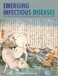
Japanese color woodcut print advertising the effectiveness of cowpox vaccine (circa 1850 A.D.)
| EID | Japanese color woodcut print advertising the effectiveness of cowpox vaccine (circa 1850 A.D.). Emerg Infect Dis. 2000;6(6):664. https://doi.org/10.3201/eid0606.ac0606 |
|---|---|
| AMA | Japanese color woodcut print advertising the effectiveness of cowpox vaccine (circa 1850 A.D.). Emerging Infectious Diseases. 2000;6(6):664. doi:10.3201/eid0606.ac0606. |
| APA | (2000). Japanese color woodcut print advertising the effectiveness of cowpox vaccine (circa 1850 A.D.). Emerging Infectious Diseases, 6(6), 664. https://doi.org/10.3201/eid0606.ac0606. |


