Perspective
Mycobacterial Aerosols and Respiratory Disease
Environmental opportunistic mycobacteria, including Mycobacterium avium, M. terrae, and the new species M. immunogenum, have been implicated in outbreaks of hypersensitivity pneumonitis or respiratory problems in a wide variety of settings. One common feature of the outbreaks has been exposure to aerosols. Aerosols have been generated from metalworking fluid during machining and grinding operations as well as from indoor swimming pools, hot tubs, and water-damaged buildings. Environmental opportunistic mycobacteria are present in drinking water, resistant to disinfection, able to provoke inflammatory reactions, and readily aerosolized. In all outbreaks, the water sources of the aerosols were disinfected. Disinfection may select for the predominance and growth of mycobacteria. Therefore, mycobacteria may be responsible, in part, for many outbreaks of hypersensitivity pneumonitis and other respiratory problems in the workplace and home.
| EID | Falkinham JO. Mycobacterial Aerosols and Respiratory Disease. Emerg Infect Dis. 2003;9(7):763-767. https://doi.org/10.3201/eid0907.020415 |
|---|---|
| AMA | Falkinham JO. Mycobacterial Aerosols and Respiratory Disease. Emerging Infectious Diseases. 2003;9(7):763-767. doi:10.3201/eid0907.020415. |
| APA | Falkinham, J. O. (2003). Mycobacterial Aerosols and Respiratory Disease. Emerging Infectious Diseases, 9(7), 763-767. https://doi.org/10.3201/eid0907.020415. |
Global Screening for Human Viral Pathogens
We propose a system for continuing surveillance of viral pathogens circulating in large human populations. We base this system on the physical isolation of viruses from large pooled samples of human serum and plasma (e.g., discarded specimens from diagnostic laboratories), followed by shotgun sequencing of the resulting genomes. The technology for concentrating virions from 100-L volumes was developed previously at Oak Ridge National Laboratory, and the means for purifying and concentrating virions from volumes in microliters have been developed recently. At the same time, marine virologists have developed efficient methods for concentrating, amplifying, and sequencing complex viral mixtures obtained from the ocean. Given this existing technology base, we believe an integrated, automated, and contained system for surveillance of the human “virome” can be implemented within 1 to 2 years. Such a system could monitor the levels of known viruses in human populations, rapidly detect outbreaks, and systematically discover novel or variant human viruses.
| EID | Anderson NG, Gerin JL, Anderson NL. Global Screening for Human Viral Pathogens. Emerg Infect Dis. 2003;9(7):768-773. https://doi.org/10.3201/eid0907.030004 |
|---|---|
| AMA | Anderson NG, Gerin JL, Anderson NL. Global Screening for Human Viral Pathogens. Emerging Infectious Diseases. 2003;9(7):768-773. doi:10.3201/eid0907.030004. |
| APA | Anderson, N. G., Gerin, J. L., & Anderson, N. L. (2003). Global Screening for Human Viral Pathogens. Emerging Infectious Diseases, 9(7), 768-773. https://doi.org/10.3201/eid0907.030004. |
Synopses
Salmonella Control Programs in Denmark
We describe Salmonella control programs of broiler chickens, layer hens, and pigs in Denmark. Major reductions in the incidence of foodborne human salmonellosis have occurred by integrated control of farms and food processing plants. Disease control has been achieved by monitoring the herds and flocks, eliminating infected animals, and diversifying animals (animals and products are processed differently depending on Salmonella status) and animal food products according to the determined risk. In 2001, the Danish society saved U.S.$25.5 million by controlling Salmonella. The total annual Salmonella control costs in year 2001 were U.S.$14.1 million (U.S.$0.075/kg of pork and U.S.$0.02/kg of broiler or egg). These costs are paid almost exclusively by the industry. The control principles described are applicable to most industrialized countries with modern intensive farming systems.
| EID | Wegener HC, Hald T, Wong LF, Madsen M, Korsgaard H, Bager F, et al. Salmonella Control Programs in Denmark. Emerg Infect Dis. 2003;9(7):774-780. https://doi.org/10.3201/eid0907.030024 |
|---|---|
| AMA | Wegener HC, Hald T, Wong LF, et al. Salmonella Control Programs in Denmark. Emerging Infectious Diseases. 2003;9(7):774-780. doi:10.3201/eid0907.030024. |
| APA | Wegener, H. C., Hald, T., Wong, L. F., Madsen, M., Korsgaard, H., Bager, F....Mølbak, K. (2003). Salmonella Control Programs in Denmark. Emerging Infectious Diseases, 9(7), 774-780. https://doi.org/10.3201/eid0907.030024. |
Disease Surveillance and the Academic, Clinical, and Public Health Communities
The Emerging Infections Programs (EIPs), a population-based network involving 10 state health departments and the Centers for Disease Control and Prevention, complement and support local, regional, and national surveillance and research efforts. EIPs depend on collaboration between public health agencies and clinical and academic institutions to perform active, population-based surveillance for infectious diseases; conduct applied epidemiologic and laboratory research; implement and evaluate pilot prevention and intervention projects; and provide capacity for flexible public health response. Recent EIP work has included monitoring the impact of a new conjugate vaccine on the epidemiology of invasive pneumococcal disease, providing the evidence base used to derive new recommendations to prevent neonatal group B streptococcal disease, measuring the impact of foodborne diseases in the United States, and developing a systematic, integrated laboratory and epidemiologic method for syndrome-based surveillance.
| EID | Pinner RW, Rebmann CA, Schuchat A, Hughes JM. Disease Surveillance and the Academic, Clinical, and Public Health Communities. Emerg Infect Dis. 2003;9(7):781-787. https://doi.org/10.3201/eid0907.030083 |
|---|---|
| AMA | Pinner RW, Rebmann CA, Schuchat A, et al. Disease Surveillance and the Academic, Clinical, and Public Health Communities. Emerging Infectious Diseases. 2003;9(7):781-787. doi:10.3201/eid0907.030083. |
| APA | Pinner, R. W., Rebmann, C. A., Schuchat, A., & Hughes, J. M. (2003). Disease Surveillance and the Academic, Clinical, and Public Health Communities. Emerging Infectious Diseases, 9(7), 781-787. https://doi.org/10.3201/eid0907.030083. |
Research
Acute Flaccid Paralysis and West Nile Virus Infection
Acute weakness associated with West Nile virus (WNV) infection has previously been attributed to a peripheral demyelinating process (Guillain-Barré syndrome); however, the exact etiology of this acute flaccid paralysis has not been systematically assessed. To thoroughly describe the clinical, laboratory, and electrodiagnostic features of this paralysis syndrome, we evaluated acute flaccid paralysis that developed in seven patients in the setting of acute WNV infection, consecutively identified in four hospitals in St. Tammany Parish and New Orleans, Louisiana, and Jackson, Mississippi. All patients had acute onset of asymmetric weakness and areflexia but no sensory abnormalities. Clinical and electrodiagnostic data suggested the involvement of spinal anterior horn cells, resulting in a poliomyelitis-like syndrome. In areas in which transmission is occurring, WNV infection should be considered in patients with acute flaccid paralysis. Recognition that such weakness may be of spinal origin may prevent inappropriate treatment and diagnostic testing.
| EID | Sejvar JJ, Leis AA, Stokic DS, Van Gerpen JA, Marfin AA, Webb R, et al. Acute Flaccid Paralysis and West Nile Virus Infection. Emerg Infect Dis. 2003;9(7):788-793. https://doi.org/10.3201/eid0907.030129 |
|---|---|
| AMA | Sejvar JJ, Leis AA, Stokic DS, et al. Acute Flaccid Paralysis and West Nile Virus Infection. Emerging Infectious Diseases. 2003;9(7):788-793. doi:10.3201/eid0907.030129. |
| APA | Sejvar, J. J., Leis, A. A., Stokic, D. S., Van Gerpen, J. A., Marfin, A. A., Webb, R....Petersen, L. R. (2003). Acute Flaccid Paralysis and West Nile Virus Infection. Emerging Infectious Diseases, 9(7), 788-793. https://doi.org/10.3201/eid0907.030129. |
West Nile Virus in Farmed Alligators
Seven alligators were submitted to the Tifton Veterinary Diagnostic and Investigational Laboratory for necropsy during two epizootics in the fall of 2001 and 2002. The alligators were raised in temperature-controlled buildings and fed a diet of horsemeat supplemented with vitamins and minerals. Histologic findings in the juvenile alligators were multiorgan necrosis, heterophilic granulomas, and heterophilic perivasculitis and were most indicative of septicemia or bacteremia. Histologic findings in a hatchling alligator were random foci of necrosis in multiple organs and mononuclear perivascular encephalitis, indicative of a viral cause. West Nile virus was isolated from submissions in 2002. Reverse transcription-polymerase chain reaction (RT-PCR) results on all submitted case samples were positive for West Nile virus for one of four cases associated with the 2001 epizootic and three of three cases associated with the 2002 epizootic. RT-PCR analysis was positive for West Nile virus in the horsemeat collected during the 2002 outbreak but negative in the horsemeat collected after the outbreak.
| EID | Miller DL, Mauel MJ, Baldwin C, Burtle G, Ingram D, Hines ME, et al. West Nile Virus in Farmed Alligators. Emerg Infect Dis. 2003;9(7):794-799. https://doi.org/10.3201/eid0907.030085 |
|---|---|
| AMA | Miller DL, Mauel MJ, Baldwin C, et al. West Nile Virus in Farmed Alligators. Emerging Infectious Diseases. 2003;9(7):794-799. doi:10.3201/eid0907.030085. |
| APA | Miller, D. L., Mauel, M. J., Baldwin, C., Burtle, G., Ingram, D., Hines, M. E....Frazier, K. S. (2003). West Nile Virus in Farmed Alligators. Emerging Infectious Diseases, 9(7), 794-799. https://doi.org/10.3201/eid0907.030085. |
Emergence and Global Spread of a Dengue Serotype 3, Subtype III Virus
Over the past two decades, dengue virus serotype 3 (DENV-3) has caused unexpected epidemics of dengue hemorrhagic fever (DHF) in Sri Lanka, East Africa, and Latin America. We used a phylogenetic approach to evaluate the roles of virus evolution and transport in the emergence of these outbreaks. Isolates from these geographically distant epidemics are closely related and belong to DENV-3, subtype III, which originated in the Indian subcontinent. The emergence of DHF in Sri Lanka in 1989 correlated with the appearance there of a new DENV-3, subtype III variant. This variant likely spread from the Indian subcontinent into Africa in the 1980s and from Africa into Latin America in the mid-1990s. DENV-3, subtype III isolates from mild and severe disease outbreaks formed genetically distinct groups, which suggests a role for viral genetics in DHF.
| EID | Messer WB, Gubler DJ, Harris E, Sivananthan K, de Silva AM. Emergence and Global Spread of a Dengue Serotype 3, Subtype III Virus. Emerg Infect Dis. 2003;9(7):800-809. https://doi.org/10.3201/eid0907.030038 |
|---|---|
| AMA | Messer WB, Gubler DJ, Harris E, et al. Emergence and Global Spread of a Dengue Serotype 3, Subtype III Virus. Emerging Infectious Diseases. 2003;9(7):800-809. doi:10.3201/eid0907.030038. |
| APA | Messer, W. B., Gubler, D. J., Harris, E., Sivananthan, K., & de Silva, A. M. (2003). Emergence and Global Spread of a Dengue Serotype 3, Subtype III Virus. Emerging Infectious Diseases, 9(7), 800-809. https://doi.org/10.3201/eid0907.030038. |
Molecular Epidemiology of O139 Vibrio cholerae: Mutation, Lateral Gene Transfer, and Founder Flush
Vibrio cholerae in O-group 139 was first isolated in 1992 and by 1993 had been found throughout the Indian subcontinent. This epidemic expansion probably resulted from a single source after a lateral gene transfer (LGT) event that changed the serotype of an epidemic V. cholerae O1 El Tor strain to O139. However, some studies found substantial genetic diversity, perhaps caused by multiple origins. To further explore the relatedness of O139 strains, we analyzed nine sequenced loci from 96 isolates from patients at the Infectious Diseases Hospital, Calcutta, from 1992 to 2000. We found 64 novel alleles distributed among 51 sequence types. LGT events produced three times the number of nucleotide changes compared to mutation. In contrast to the traditional concept of epidemic spread of a homogeneous clone, the establishment of variant alleles generated by LGT during the rapid expansion of a clonal bacterial population may be a paradigm in infections and epidemics.
| EID | Garg P, Aydanian A, Smith DW, Morris J, Nair GB, Stine O. Molecular Epidemiology of O139 Vibrio cholerae: Mutation, Lateral Gene Transfer, and Founder Flush. Emerg Infect Dis. 2003;9(7):810-814. https://doi.org/10.3201/eid0907.020760 |
|---|---|
| AMA | Garg P, Aydanian A, Smith DW, et al. Molecular Epidemiology of O139 Vibrio cholerae: Mutation, Lateral Gene Transfer, and Founder Flush. Emerging Infectious Diseases. 2003;9(7):810-814. doi:10.3201/eid0907.020760. |
| APA | Garg, P., Aydanian, A., Smith, D. W., Morris, J., Nair, G. B., & Stine, O. (2003). Molecular Epidemiology of O139 Vibrio cholerae: Mutation, Lateral Gene Transfer, and Founder Flush. Emerging Infectious Diseases, 9(7), 810-814. https://doi.org/10.3201/eid0907.020760. |
Amoeba-Resisting Bacteria and Ventilator-Associated Pneumonia
To evaluate the role of amoeba-associated bacteria as agents of ventilator-associated pneumonia (VAP), we tested the water from an intensive care unit (ICU) every week for 6 months for such bacteria isolates; serum samples and bronchoalveolar lavage samples (BAL) were also obtained from 30 ICU patients. BAL samples were examined for amoeba-associated bacteria DNA by suicide-polymerase chain reaction, and serum samples were tested against ICU amoeba-associated bacteria. A total of 310 amoeba-associated bacteria from10 species were isolated. Twelve of 30 serum samples seroconverted to one amoeba-associated bacterium isolated in the ICU, mainly Legionella anisa and Bosea massiliensis, the most common isolates from water (p=0.021). Amoeba-associated bacteria DNA was detected in BAL samples from two patients whose samples later seroconverted. Seroconversion was significantly associated with VAP and systemic inflammatory response syndrome, especially in patients for whom no etiologic agent was found by usual microbiologic investigations. Amoeba-associated bacteria might be a cause of VAP in ICUs, especially when microbiologic investigations are negative.
| EID | La Scola B, Boyadjiev I, Greub G, Khamis A, Martin C, Raoult D. Amoeba-Resisting Bacteria and Ventilator-Associated Pneumonia. Emerg Infect Dis. 2003;9(7):815-821. https://doi.org/10.3201/eid0907.030065 |
|---|---|
| AMA | La Scola B, Boyadjiev I, Greub G, et al. Amoeba-Resisting Bacteria and Ventilator-Associated Pneumonia. Emerging Infectious Diseases. 2003;9(7):815-821. doi:10.3201/eid0907.030065. |
| APA | La Scola, B., Boyadjiev, I., Greub, G., Khamis, A., Martin, C., & Raoult, D. (2003). Amoeba-Resisting Bacteria and Ventilator-Associated Pneumonia. Emerging Infectious Diseases, 9(7), 815-821. https://doi.org/10.3201/eid0907.030065. |
Antimicrobial Resistance Markers of Class 1 and Class 2 Integron-bearing Escherichia coli from Irrigation Water and Sediments
Municipal and agricultural pollution affects the Rio Grande, a river that separates the United States from Mexico. Three hundred and twenty-two Escherichia coli isolates were examined for multiple antibiotic resistance phenotypes and the prevalence of class 1 and class 2 integron sequences. Thirty-two (10%) of the isolates were resistant to multiple antibiotics. Four (13%) of these isolates contained class 1–specific integron sequences; one isolate contained class 2 integron–specific sequences. Sequencing showed that the class 1 integron–bearing strain contained two distinct gene cassettes, sat-1 and aadA. Although three of the four class 1 integron–bearing strains harbored the aadA sequence, none of the strains was phenotypically resistant to streptomycin. These results suggest that integron-bearing E. coli strains can be present in contaminated irrigation canals and that these isolates may not express these resistance markers.
| EID | Roe MT, Vega E, Pillai SD. Antimicrobial Resistance Markers of Class 1 and Class 2 Integron-bearing Escherichia coli from Irrigation Water and Sediments. Emerg Infect Dis. 2003;9(7):822-826. https://doi.org/10.3201/eid0907.020529 |
|---|---|
| AMA | Roe MT, Vega E, Pillai SD. Antimicrobial Resistance Markers of Class 1 and Class 2 Integron-bearing Escherichia coli from Irrigation Water and Sediments. Emerging Infectious Diseases. 2003;9(7):822-826. doi:10.3201/eid0907.020529. |
| APA | Roe, M. T., Vega, E., & Pillai, S. D. (2003). Antimicrobial Resistance Markers of Class 1 and Class 2 Integron-bearing Escherichia coli from Irrigation Water and Sediments. Emerging Infectious Diseases, 9(7), 822-826. https://doi.org/10.3201/eid0907.020529. |
Hantavirus Prevalence in the IX Region of Chile
An epidemiologic and seroprevalence survey was conducted (n=830) to assess proportion of persons exposed to hantavirus in IX Region Chile, which accounts for 25% of reported cases of hantavirus cardiopulmonary syndrome. This region has three geographic areas with different disease incidences and a high proportion of aboriginals. Serum samples were tested for immunoglobulin (Ig) G antibodies by enzyme-linked immunosorbent assay against Sin Nombre virus N antigen by strip immunoblot assay against Sin Nombre, Puumala, Río Mamoré, and Seoul N antigens. Samples from six patients were positive for IgG antibodies reactive with Andes virus; all patients lived in the Andes Mountains. Foresting was also associated with seropositivity; but not sex, age, race, rodent exposure, or farming activities. Exposure to hantavirus varies in different communities of IX Region. Absence of history of pneumonia or hospital admission in persons with specific IgG antibodies suggests that infection is clinically inapparent.
| EID | Frey MT, Vial PC, Castillo CH, Godoy PM, Hjelle B, Ferrés MG. Hantavirus Prevalence in the IX Region of Chile. Emerg Infect Dis. 2003;9(7):827-832. https://doi.org/10.3201/eid0907.020587 |
|---|---|
| AMA | Frey MT, Vial PC, Castillo CH, et al. Hantavirus Prevalence in the IX Region of Chile. Emerging Infectious Diseases. 2003;9(7):827-832. doi:10.3201/eid0907.020587. |
| APA | Frey, M. T., Vial, P. C., Castillo, C. H., Godoy, P. M., Hjelle, B., & Ferrés, M. G. (2003). Hantavirus Prevalence in the IX Region of Chile. Emerging Infectious Diseases, 9(7), 827-832. https://doi.org/10.3201/eid0907.020587. |
Antimicrobial Susceptibility Breakpoints and First-Step parC Mutations in Streptococcus pneumoniae: Redefining Fluoroquinolone Resistance
Clinical antimicrobial susceptibility breakpoints are used to predict the clinical outcome of antimicrobial treatment. In contrast, microbiologic breakpoints are used to identify isolates that may be categorized as susceptible when applying clinical breakpoints but harbor resistance mechanisms that result in their reduced susceptibility to the agent being tested. Currently, the National Committee for Clinical Laboratory Standards (NCCLS) guidelines utilize clinical breakpoints to characterize the activity of the fluoroquinolones against Streptococcus pneumoniae. To determine whether levofloxacin breakpoints can identify isolates that harbor recognized resistance mechanisms, we examined 115 S. pneumoniae isolates with a levofloxacin MIC of >2 μg/mL for first-step parC mutations. A total of 48 (59%) of 82 isolates with a levofloxacin MIC of 2 μg/mL, a level considered susceptible by NCCLS criteria, had a first-step mutation in parC. Whether surveillance programs that use levofloxacin data can effectively detect emerging resistance and whether fluoroquinolones can effectively treat infections caused by such isolates should be evaluated.
| EID | Lim S, Bast D, McGeer A, de Azavedo J, Low DE. Antimicrobial Susceptibility Breakpoints and First-Step parC Mutations in Streptococcus pneumoniae: Redefining Fluoroquinolone Resistance. Emerg Infect Dis. 2003;9(7):833-837. https://doi.org/10.3201/eid0907.020589 |
|---|---|
| AMA | Lim S, Bast D, McGeer A, et al. Antimicrobial Susceptibility Breakpoints and First-Step parC Mutations in Streptococcus pneumoniae: Redefining Fluoroquinolone Resistance. Emerging Infectious Diseases. 2003;9(7):833-837. doi:10.3201/eid0907.020589. |
| APA | Lim, S., Bast, D., McGeer, A., de Azavedo, J., & Low, D. E. (2003). Antimicrobial Susceptibility Breakpoints and First-Step parC Mutations in Streptococcus pneumoniae: Redefining Fluoroquinolone Resistance. Emerging Infectious Diseases, 9(7), 833-837. https://doi.org/10.3201/eid0907.020589. |
Mutations in Putative Mutator Genes of Mycobacterium tuberculosis Strains of the W-Beijing Family
Alterations in genes involved in the repair of DNA mutations (mut genes) result in an increased mutation frequency and better adaptability of the bacterium to stressful conditions. W-Beijing genotype strains displayed unique missense alterations in three putative mut genes, including two of the mutT type (Rv3908 and mutT2) and ogt. These polymorphisms were found to be characteristic and unique to W-Beijing phylogenetic lineage. Analysis of the mut genes in 55 representative W-Beijing isolates suggests a sequential acquisition of the mutations, elucidating a plausible pathway of the molecular evolution of this clonal family. The acquisition of mut genes may explain in part the ability of the isolates of W-Beijing type to rapidly adapt to their environment.
| EID | Rad ME, Bifani P, Martin C, Kremer K, Samper S, Rauzier J, et al. Mutations in Putative Mutator Genes of Mycobacterium tuberculosis Strains of the W-Beijing Family. Emerg Infect Dis. 2003;9(7):838-845. https://doi.org/10.3201/eid0907.020803 |
|---|---|
| AMA | Rad ME, Bifani P, Martin C, et al. Mutations in Putative Mutator Genes of Mycobacterium tuberculosis Strains of the W-Beijing Family. Emerging Infectious Diseases. 2003;9(7):838-845. doi:10.3201/eid0907.020803. |
| APA | Rad, M. E., Bifani, P., Martin, C., Kremer, K., Samper, S., Rauzier, J....Gicquel, B. (2003). Mutations in Putative Mutator Genes of Mycobacterium tuberculosis Strains of the W-Beijing Family. Emerging Infectious Diseases, 9(7), 838-845. https://doi.org/10.3201/eid0907.020803. |
Yellow Pygmy Rice Rat (Oligoryzomys flavescens) and Hantavirus Pulmonary Syndrome in Uruguay
During 5,230 trapping nights, 672 small mammals were trapped in the areas where most hantavirus pulmonary syndrome (HPS) cases occur in Uruguay. Yellow pygmy rice rats (Oligoryzomys flavescens) were the only rodents that showed evidence of antibodies to hantavirus, with a seroprevalence of 2.6%. The rodents were trapped in all the explored environments, and most of the seropositive rodents were found in habitats frequented by humans. Nucleotide sequences were obtained from four HPS case-patients and four yellow pygmy rice rats of the M genome segment. Sequence comparison and phylogenetic analysis showed that rodent-borne viruses and viruses from three HPS case-patients form a well-supported clade and share a 96.4% identity with the previously characterized Central Plata hantavirus. These results suggest that yellow pygmy rice rat (O. flavescens) may be the host for Central Plata, a hantavirus associated with HPS in the southern area of Uruguay.[
| EID | Delfraro A, Clara M, Tomé L, Achaval F, Levis S, Calderón G, et al. Yellow Pygmy Rice Rat (Oligoryzomys flavescens) and Hantavirus Pulmonary Syndrome in Uruguay. Emerg Infect Dis. 2003;9(7):846-852. https://doi.org/10.3201/eid0907.030044 |
|---|---|
| AMA | Delfraro A, Clara M, Tomé L, et al. Yellow Pygmy Rice Rat (Oligoryzomys flavescens) and Hantavirus Pulmonary Syndrome in Uruguay. Emerging Infectious Diseases. 2003;9(7):846-852. doi:10.3201/eid0907.030044. |
| APA | Delfraro, A., Clara, M., Tomé, L., Achaval, F., Levis, S., Calderón, G....Arbiza, J. (2003). Yellow Pygmy Rice Rat (Oligoryzomys flavescens) and Hantavirus Pulmonary Syndrome in Uruguay. Emerging Infectious Diseases, 9(7), 846-852. https://doi.org/10.3201/eid0907.030044. |
Dispatches
Serologic Evidence of West Nile Virus Infection in Horses, Coahuila State, Mexico
Serum samples were obtained from 24 horses in the State of Coahuila, Mexico, in December 2002. Antibodies to West Nile virus were detected by epitope-blocking enzyme-linked immunosorbent assay and confirmed by plaque reduction neutralization test in 15 (62.5%) horses. We report the first West Nile virus activity in northern Mexico.
| EID | Blitvich BJ, Fernandez-Salas I, Contreras-Cordero JF, Marlenee NL, Gonzalez-Rojas JI, Komar N, et al. Serologic Evidence of West Nile Virus Infection in Horses, Coahuila State, Mexico. Emerg Infect Dis. 2003;9(7):853-856. https://doi.org/10.3201/eid0907.030166 |
|---|---|
| AMA | Blitvich BJ, Fernandez-Salas I, Contreras-Cordero JF, et al. Serologic Evidence of West Nile Virus Infection in Horses, Coahuila State, Mexico. Emerging Infectious Diseases. 2003;9(7):853-856. doi:10.3201/eid0907.030166. |
| APA | Blitvich, B. J., Fernandez-Salas, I., Contreras-Cordero, J. F., Marlenee, N. L., Gonzalez-Rojas, J. I., Komar, N....Beaty, B. J. (2003). Serologic Evidence of West Nile Virus Infection in Horses, Coahuila State, Mexico. Emerging Infectious Diseases, 9(7), 853-856. https://doi.org/10.3201/eid0907.030166. |
Serologic Evidence of West Nile Virus Infection in Horses, Yucatan State, Mexico
Serum samples were obtained from 252 horses in the State of Yucatan, Mexico, from July to October 2002. Antibodies to West Nile virus were detected by epitope-blocking enzyme-linked immunosorbent assays in three (1.2%) horses and confirmed by plaque reduction neutralization test. We report the first West Nile virus activity in the State of Yucatan.
| EID | Loroño-Pino MA, Blitvich BJ, Farfán-Ale JA, Puerto FI, Blanco JM, Marlenee NL, et al. Serologic Evidence of West Nile Virus Infection in Horses, Yucatan State, Mexico. Emerg Infect Dis. 2003;9(7):857-859. https://doi.org/10.3201/eid0907.030167 |
|---|---|
| AMA | Loroño-Pino MA, Blitvich BJ, Farfán-Ale JA, et al. Serologic Evidence of West Nile Virus Infection in Horses, Yucatan State, Mexico. Emerging Infectious Diseases. 2003;9(7):857-859. doi:10.3201/eid0907.030167. |
| APA | Loroño-Pino, M. A., Blitvich, B. J., Farfán-Ale, J. A., Puerto, F. I., Blanco, J. M., Marlenee, N. L....Beaty, B. J. (2003). Serologic Evidence of West Nile Virus Infection in Horses, Yucatan State, Mexico. Emerging Infectious Diseases, 9(7), 857-859. https://doi.org/10.3201/eid0907.030167. |
Serologic Evidence of West Nile Virus Transmission, Jamaica, West Indies
In spring 2002, an intensive avian serosurvey was initiated in Jamaica, Puerto Rico, and Mexico. We collected >1,600 specimens from resident and nonresident neotropical migratory birds before their northerly migrations. Plaque reduction neutralization test results indicated specific neutralizing antibodies to West Nile virus in 11 resident species from Jamaica.
| EID | Dupuis AP, Marra PP, Kramer LD. Serologic Evidence of West Nile Virus Transmission, Jamaica, West Indies. Emerg Infect Dis. 2003;9(7):860-863. https://doi.org/10.3201/eid0907.030249 |
|---|---|
| AMA | Dupuis AP, Marra PP, Kramer LD. Serologic Evidence of West Nile Virus Transmission, Jamaica, West Indies. Emerging Infectious Diseases. 2003;9(7):860-863. doi:10.3201/eid0907.030249. |
| APA | Dupuis, A. P., Marra, P. P., & Kramer, L. D. (2003). Serologic Evidence of West Nile Virus Transmission, Jamaica, West Indies. Emerging Infectious Diseases, 9(7), 860-863. https://doi.org/10.3201/eid0907.030249. |
Sulfa Resistance and Dihydropteroate Synthase Mutants in Recurrent Pneumocystis carinii Pneumonia
Failure of sulfa or sulfone prophylaxis is associated with mutations in Pneumocystis carinii gene coding for dihydropteroate synthase (DHPS). The DHPS genotype was analyzed in AIDS patients who had two separate episodes of P. carinii pneumonia. The results suggest that DHPS mutations can be selected de novo within patients by the pressure of a sulfa or sulfone drug.
| EID | Nahimana A, Rabodonirina M, Helweg-Larsen J, Meneau I, Francioli P, Bille J, et al. Sulfa Resistance and Dihydropteroate Synthase Mutants in Recurrent Pneumocystis carinii Pneumonia. Emerg Infect Dis. 2003;9(7):864-867. https://doi.org/10.3201/eid0907.020753 |
|---|---|
| AMA | Nahimana A, Rabodonirina M, Helweg-Larsen J, et al. Sulfa Resistance and Dihydropteroate Synthase Mutants in Recurrent Pneumocystis carinii Pneumonia. Emerging Infectious Diseases. 2003;9(7):864-867. doi:10.3201/eid0907.020753. |
| APA | Nahimana, A., Rabodonirina, M., Helweg-Larsen, J., Meneau, I., Francioli, P., Bille, J....Hauser, P. M. (2003). Sulfa Resistance and Dihydropteroate Synthase Mutants in Recurrent Pneumocystis carinii Pneumonia. Emerging Infectious Diseases, 9(7), 864-867. https://doi.org/10.3201/eid0907.020753. |
VIM- and IMP-Type Metallo-β-lactamase–Producing Pseudomonas spp. and Acinetobacter spp. in Korean Hospitals
We determined the occurrence of acquired metallo-β-lactamase (MBL)–producing bacteria in Korean hospitals. Among the isolates nonsusceptible to imipenem that were collected from 28 hospitals from 2000 to 2001, 44 (11.4%) of 387 Pseudomonas spp. and 38 (14.2%) of 267 Acinetobacter spp. infections produced MBL and had alleles of blaVIM-2 or blaIMP-1. MBL-producing isolates were detected in 60.7% of the hospitals.
| EID | Lee K, Lee WG, Uh Y, Ha GY, Cho J, Chong Y. VIM- and IMP-Type Metallo-β-lactamase–Producing Pseudomonas spp. and Acinetobacter spp. in Korean Hospitals. Emerg Infect Dis. 2003;9(7):868-871. https://doi.org/10.3201/eid0907.030012 |
|---|---|
| AMA | Lee K, Lee WG, Uh Y, et al. VIM- and IMP-Type Metallo-β-lactamase–Producing Pseudomonas spp. and Acinetobacter spp. in Korean Hospitals. Emerging Infectious Diseases. 2003;9(7):868-871. doi:10.3201/eid0907.030012. |
| APA | Lee, K., Lee, W. G., Uh, Y., Ha, G. Y., Cho, J., & Chong, Y. (2003). VIM- and IMP-Type Metallo-β-lactamase–Producing Pseudomonas spp. and Acinetobacter spp. in Korean Hospitals. Emerging Infectious Diseases, 9(7), 868-871. https://doi.org/10.3201/eid0907.030012. |
Leishmaniasis in Germany
In 2000, a reference center was created to systematically record leishmaniases in Germany. We analyzed 58 cases of leishmaniases imported during a 2-year period. These findings will serve as a baseline for the sandfly vector’s anticipated northward move because of global warming and as an advisory for immunocompromised persons traveling to leishmaniasis-endemic areas.
| EID | Harms G, Schönian G, Feldmeier H. Leishmaniasis in Germany. Emerg Infect Dis. 2003;9(7):872-875. https://doi.org/10.3201/eid0907.030023 |
|---|---|
| AMA | Harms G, Schönian G, Feldmeier H. Leishmaniasis in Germany. Emerging Infectious Diseases. 2003;9(7):872-875. doi:10.3201/eid0907.030023. |
| APA | Harms, G., Schönian, G., & Feldmeier, H. (2003). Leishmaniasis in Germany. Emerging Infectious Diseases, 9(7), 872-875. https://doi.org/10.3201/eid0907.030023. |
Probable Dengue Virus Infection Among Italian Troops, East Timor, 1999–2000
To investigate the attack rate and risk factors for probable dengue fever, a cross-sectional study was conducted of an Italian military unit after its deployment to East Timor. Probable dengue was contracted by 16 (6.6%) of 241 army troops and caused half of all medical evacuations (12/24); no cases were detected among navy and air force personnel.
| EID | Peragallo MS, Nicoletti L, Lista F, D’Amelio R. Probable Dengue Virus Infection Among Italian Troops, East Timor, 1999–2000. Emerg Infect Dis. 2003;9(7):876-880. https://doi.org/10.3201/eid0907.020496 |
|---|---|
| AMA | Peragallo MS, Nicoletti L, Lista F, et al. Probable Dengue Virus Infection Among Italian Troops, East Timor, 1999–2000. Emerging Infectious Diseases. 2003;9(7):876-880. doi:10.3201/eid0907.020496. |
| APA | Peragallo, M. S., Nicoletti, L., Lista, F., & D’Amelio, R. (2003). Probable Dengue Virus Infection Among Italian Troops, East Timor, 1999–2000. Emerging Infectious Diseases, 9(7), 876-880. https://doi.org/10.3201/eid0907.020496. |
HIV Epidemic among Young Thai Men, 1991–2000
Characterization of the HIV epidemic in Thailand has benefited from the systematic testing of young men upon entry into the military. These data, which have shown that public health measures can reverse an HIV epidemic, have been reanalyzed with current geographic information systems methods. The resulting maps, thus far, are the best means of visualizing the geography of the dynamic HIV epidemic in Thailand.
| EID | Torugsa K, Anderson S, Thongsen N, Sirisopana N, Jugsudee A, Junlananto P, et al. HIV Epidemic among Young Thai Men, 1991–2000. Emerg Infect Dis. 2003;9(7):881-883. https://doi.org/10.3201/eid0907.020653 |
|---|---|
| AMA | Torugsa K, Anderson S, Thongsen N, et al. HIV Epidemic among Young Thai Men, 1991–2000. Emerging Infectious Diseases. 2003;9(7):881-883. doi:10.3201/eid0907.020653. |
| APA | Torugsa, K., Anderson, S., Thongsen, N., Sirisopana, N., Jugsudee, A., Junlananto, P....Brown, A. E. (2003). HIV Epidemic among Young Thai Men, 1991–2000. Emerging Infectious Diseases, 9(7), 881-883. https://doi.org/10.3201/eid0907.020653. |
Letters
Taenia solium Cysticercosis, Irian Jaya, Indonesia
| EID | Wandra T, Ito A, Yamasaki H, Suroso T, Margono SS. Taenia solium Cysticercosis, Irian Jaya, Indonesia. Emerg Infect Dis. 2003;9(7):884-885. https://doi.org/10.3201/eid0907.020709 |
|---|---|
| AMA | Wandra T, Ito A, Yamasaki H, et al. Taenia solium Cysticercosis, Irian Jaya, Indonesia. Emerging Infectious Diseases. 2003;9(7):884-885. doi:10.3201/eid0907.020709. |
| APA | Wandra, T., Ito, A., Yamasaki, H., Suroso, T., & Margono, S. S. (2003). Taenia solium Cysticercosis, Irian Jaya, Indonesia. Emerging Infectious Diseases, 9(7), 884-885. https://doi.org/10.3201/eid0907.020709. |
Recombinant Vaccine–Derived Poliovirus in Madagascar
| EID | Rousset D, Rakoto-Andrianarivelo M, Razafindratsimandresy R, Randriamanalina B, Guillot S, Balanant J, et al. Recombinant Vaccine–Derived Poliovirus in Madagascar. Emerg Infect Dis. 2003;9(7):885-887. https://doi.org/10.3201/eid0907.020692 |
|---|---|
| AMA | Rousset D, Rakoto-Andrianarivelo M, Razafindratsimandresy R, et al. Recombinant Vaccine–Derived Poliovirus in Madagascar. Emerging Infectious Diseases. 2003;9(7):885-887. doi:10.3201/eid0907.020692. |
| APA | Rousset, D., Rakoto-Andrianarivelo, M., Razafindratsimandresy, R., Randriamanalina, B., Guillot, S., Balanant, J....Delpeyroux, F. (2003). Recombinant Vaccine–Derived Poliovirus in Madagascar. Emerging Infectious Diseases, 9(7), 885-887. https://doi.org/10.3201/eid0907.020692. |
West Nile Virus Infection in Crocodiles
| EID | Steinman A, Banet-Noach C, Tal S, Levi O, Simanov L, Perk S, et al. West Nile Virus Infection in Crocodiles. Emerg Infect Dis. 2003;9(7):887-889. https://doi.org/10.3201/eid0907.020816 |
|---|---|
| AMA | Steinman A, Banet-Noach C, Tal S, et al. West Nile Virus Infection in Crocodiles. Emerging Infectious Diseases. 2003;9(7):887-889. doi:10.3201/eid0907.020816. |
| APA | Steinman, A., Banet-Noach, C., Tal, S., Levi, O., Simanov, L., Perk, S....Shpigel, N. (2003). West Nile Virus Infection in Crocodiles. Emerging Infectious Diseases, 9(7), 887-889. https://doi.org/10.3201/eid0907.020816. |
Rickettsia aeschlimannii in Spain: Molecular Evidence in Hyalomma marginatum and Five Other Tick Species that Feed on Humans
| EID | Rickettsia aeschlimannii in Spain: Molecular Evidence in Hyalomma marginatum and Five Other Tick Species that Feed on Humans. Emerg Infect Dis. 2003;9(7):889-890. https://doi.org/10.3201/eid0907.030077 |
|---|---|
| AMA | Rickettsia aeschlimannii in Spain: Molecular Evidence in Hyalomma marginatum and Five Other Tick Species that Feed on Humans. Emerging Infectious Diseases. 2003;9(7):889-890. doi:10.3201/eid0907.030077. |
| APA | (2003). Rickettsia aeschlimannii in Spain: Molecular Evidence in Hyalomma marginatum and Five Other Tick Species that Feed on Humans. Emerging Infectious Diseases, 9(7), 889-890. https://doi.org/10.3201/eid0907.030077. |
Hantaviruses in São Paulo State, Brazil
| EID | Figueiredo LT, Moreli ML, Campos GM, Sousa RL. Hantaviruses in São Paulo State, Brazil. Emerg Infect Dis. 2003;9(7):891-892. https://doi.org/10.3201/eid0907.030087 |
|---|---|
| AMA | Figueiredo LT, Moreli ML, Campos GM, et al. Hantaviruses in São Paulo State, Brazil. Emerging Infectious Diseases. 2003;9(7):891-892. doi:10.3201/eid0907.030087. |
| APA | Figueiredo, L. T., Moreli, M. L., Campos, G. M., & Sousa, R. L. (2003). Hantaviruses in São Paulo State, Brazil. Emerging Infectious Diseases, 9(7), 891-892. https://doi.org/10.3201/eid0907.030087. |
Israeli Spotted Fever Rickettsia in Sicilian Rhipicephalus sanguineus Ticks
| EID | Giammanco GM, Mansueto S, Ammatuna P, Vitale G. Israeli Spotted Fever Rickettsia in Sicilian Rhipicephalus sanguineus Ticks. Emerg Infect Dis. 2003;9(7):892-893. https://doi.org/10.3201/eid0907.030109 |
|---|---|
| AMA | Giammanco GM, Mansueto S, Ammatuna P, et al. Israeli Spotted Fever Rickettsia in Sicilian Rhipicephalus sanguineus Ticks. Emerging Infectious Diseases. 2003;9(7):892-893. doi:10.3201/eid0907.030109. |
| APA | Giammanco, G. M., Mansueto, S., Ammatuna, P., & Vitale, G. (2003). Israeli Spotted Fever Rickettsia in Sicilian Rhipicephalus sanguineus Ticks. Emerging Infectious Diseases, 9(7), 892-893. https://doi.org/10.3201/eid0907.030109. |
Co-feeding Transmission and Its Contribution to the Perpetuation of the Lyme Disease Spirochete Borrelia afzelii (In Reply)
| EID | Randolph S, Gern L. Co-feeding Transmission and Its Contribution to the Perpetuation of the Lyme Disease Spirochete Borrelia afzelii (In Reply). Emerg Infect Dis. 2003;9(7):893-894. https://doi.org/10.3201/eid0907.030116 |
|---|---|
| AMA | Randolph S, Gern L. Co-feeding Transmission and Its Contribution to the Perpetuation of the Lyme Disease Spirochete Borrelia afzelii (In Reply). Emerging Infectious Diseases. 2003;9(7):893-894. doi:10.3201/eid0907.030116. |
| APA | Randolph, S., & Gern, L. (2003). Co-feeding Transmission and Its Contribution to the Perpetuation of the Lyme Disease Spirochete Borrelia afzelii (In Reply). Emerging Infectious Diseases, 9(7), 893-894. https://doi.org/10.3201/eid0907.030116. |
Co-feeding Transmission and Its Contribution to the Perpetuation of the Lyme Disease Spirochete Borrelia afzelii (In Reply to Randolph and Gern)
| EID | Richter D, Allgöwer R, Matuschka F. Co-feeding Transmission and Its Contribution to the Perpetuation of the Lyme Disease Spirochete Borrelia afzelii (In Reply to Randolph and Gern). Emerg Infect Dis. 2003;9(7):895-896. https://doi.org/10.3201/eid0907.030342 |
|---|---|
| AMA | Richter D, Allgöwer R, Matuschka F. Co-feeding Transmission and Its Contribution to the Perpetuation of the Lyme Disease Spirochete Borrelia afzelii (In Reply to Randolph and Gern). Emerging Infectious Diseases. 2003;9(7):895-896. doi:10.3201/eid0907.030342. |
| APA | Richter, D., Allgöwer, R., & Matuschka, F. (2003). Co-feeding Transmission and Its Contribution to the Perpetuation of the Lyme Disease Spirochete Borrelia afzelii (In Reply to Randolph and Gern). Emerging Infectious Diseases, 9(7), 895-896. https://doi.org/10.3201/eid0907.030342. |
Another Dimension
Dejà Vu
| EID | Artinian BL. Dejà Vu. Emerg Infect Dis. 2003;9(7):888. https://doi.org/10.3201/eid0907.ad0907 |
|---|---|
| AMA | Artinian BL. Dejà Vu. Emerging Infectious Diseases. 2003;9(7):888. doi:10.3201/eid0907.ad0907. |
| APA | Artinian, B. L. (2003). Dejà Vu. Emerging Infectious Diseases, 9(7), 888. https://doi.org/10.3201/eid0907.ad0907. |
About the Cover
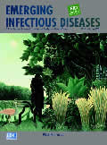
Henri Rousseau—known as Le Douanier Rousseau (1844–1910). The Snake Charmer (1907).
| EID | Potter P. Henri Rousseau—known as Le Douanier Rousseau (1844–1910). The Snake Charmer (1907).. Emerg Infect Dis. 2003;9(7):901. https://doi.org/10.3201/eid0907.ac0907 |
|---|---|
| AMA | Potter P. Henri Rousseau—known as Le Douanier Rousseau (1844–1910). The Snake Charmer (1907).. Emerging Infectious Diseases. 2003;9(7):901. doi:10.3201/eid0907.ac0907. |
| APA | Potter, P. (2003). Henri Rousseau—known as Le Douanier Rousseau (1844–1910). The Snake Charmer (1907).. Emerging Infectious Diseases, 9(7), 901. https://doi.org/10.3201/eid0907.ac0907. |
News and Notes
West Nile Virus Southeast Conference
| EID | Rimland D, Koplan J, Stephens DS. West Nile Virus Southeast Conference. Emerg Infect Dis. 2003;9(7):897-898. https://doi.org/10.3201/eid0907.030254 |
|---|---|
| AMA | Rimland D, Koplan J, Stephens DS. West Nile Virus Southeast Conference. Emerging Infectious Diseases. 2003;9(7):897-898. doi:10.3201/eid0907.030254. |
| APA | Rimland, D., Koplan, J., & Stephens, D. S. (2003). West Nile Virus Southeast Conference. Emerging Infectious Diseases, 9(7), 897-898. https://doi.org/10.3201/eid0907.030254. |
West Nile Virus and Wildlife Health
| EID | Marra PP, Griffing SM, McLean RG. West Nile Virus and Wildlife Health. Emerg Infect Dis. 2003;9(7):898-899. https://doi.org/10.3201/eid0907.030277 |
|---|---|
| AMA | Marra PP, Griffing SM, McLean RG. West Nile Virus and Wildlife Health. Emerging Infectious Diseases. 2003;9(7):898-899. doi:10.3201/eid0907.030277. |
| APA | Marra, P. P., Griffing, S. M., & McLean, R. G. (2003). West Nile Virus and Wildlife Health. Emerging Infectious Diseases, 9(7), 898-899. https://doi.org/10.3201/eid0907.030277. |


