Articles from Emerging Infectious Diseases
Volume 31, Number 5—May 2025
Synopses
Outbreak of Marburg Virus Disease, Equatorial Guinea, 2023
In February 2023, the government of Equatorial Guinea declared an outbreak of Marburg virus disease. We describe the response structure and epidemiologic characteristics, including case-patient demographics, clinical manifestations, risk factors, and the serial interval and timing of symptom onset, treatment seeking, and recovery or death. We identified 16 laboratory-confirmed and 23 probable cases of Marburg virus disease in 5 districts and noted several unlinked chains of transmission and a case-fatality ratio of 90% (35/39 cases). Transmission was concentrated in family clusters and healthcare settings. The median serial interval was 18.5 days; most transmission occurred during late-stage disease. Rapid isolation of symptomatic case-patients is critical in preventing transmission and improving patient outcomes; community engagement and surveillance strengthening should be prioritized in emerging outbreaks. Further analysis of this outbreak and a One Health surveillance approach can help prevent and prepare for future potential spillover events.
| EID | Ngai S, Evers E, Seoane A, Ameh G, Anoko JN, Barnadas C, et al. Outbreak of Marburg Virus Disease, Equatorial Guinea, 2023. Emerg Infect Dis. 2025;31(5):887-895. https://doi.org/10.3201/eid3105.241749 |
|---|---|
| AMA | Ngai S, Evers E, Seoane A, et al. Outbreak of Marburg Virus Disease, Equatorial Guinea, 2023. Emerging Infectious Diseases. 2025;31(5):887-895. doi:10.3201/eid3105.241749. |
| APA | Ngai, S., Evers, E., Seoane, A., Ameh, G., Anoko, J. N., Barnadas, C....Ndoho, F. (2025). Outbreak of Marburg Virus Disease, Equatorial Guinea, 2023. Emerging Infectious Diseases, 31(5), 887-895. https://doi.org/10.3201/eid3105.241749. |
Comprehensive Survival Analysis of Alveolar Echinococcosis Patients, University Hospital Zurich, Zurich, Switzerland, 1973–2022
Alveolar echinococcosis (AE) is a zoonotic disease of increasing concern worldwide. Before benzimidazole drug therapy, 10-year death rates were 90% without surgical resection. In unresectable patients, long-term benzimidazole therapy is highly effective in stabilizing the disease course. We performed a retrospective study of 334 AE patients treated at the University Hospital Zurich, Zurich, Switzerland, during 1973–2022. Annual diagnoses increased over time, and more cases were detected by chance at earlier stages. Ninety patients died, mostly from causes unrelated to AE. Relative survival of AE patients compared with the population of Switzerland demonstrated a steady decrease 5 years after diagnosis. Patient age at diagnosis was the primary variable associated with overall survival. In a propensity-score matched survival analysis, early curative surgery was associated with overall improvement but not AE-specific survival. We conclude that survival of patients with AE is limited by non-AE causes and that early curative surgery does not improve AE-specific survival.
| EID | Deibel A, Kindler Y, Mita R, Ghafoor S, Meyer zu Schwabedissen C, Brunner-Geissmann B, et al. Comprehensive Survival Analysis of Alveolar Echinococcosis Patients, University Hospital Zurich, Zurich, Switzerland, 1973–2022. Emerg Infect Dis. 2025;31(5):906-916. https://doi.org/10.3201/eid3105.241608 |
|---|---|
| AMA | Deibel A, Kindler Y, Mita R, et al. Comprehensive Survival Analysis of Alveolar Echinococcosis Patients, University Hospital Zurich, Zurich, Switzerland, 1973–2022. Emerging Infectious Diseases. 2025;31(5):906-916. doi:10.3201/eid3105.241608. |
| APA | Deibel, A., Kindler, Y., Mita, R., Ghafoor, S., Meyer zu Schwabedissen, C., Brunner-Geissmann, B....Müllhaupt, B. (2025). Comprehensive Survival Analysis of Alveolar Echinococcosis Patients, University Hospital Zurich, Zurich, Switzerland, 1973–2022. Emerging Infectious Diseases, 31(5), 906-916. https://doi.org/10.3201/eid3105.241608. |
Invasive aspergillosis (IA) caused by Aspergillus flavus remains poorly described. We retrospectively analyzed 54 cases of IA caused by A. flavus reported in France during 2012–2018. Among cases, underlying IA risk factors were malignancy, solid organ transplantation, and diabetes. Most (87%, 47/54) infections were localized, of which 33 were pleuropulmonary and 13 were ear-nose-throat (ENT) infection sites. Malignancy (70% [23/33]) and solid organ transplantation (21% [7/33]) were the main risk factors in localized pulmonary infections, and diabetes mellitus was associated with localized ENT involvement (61.5%, [8/13]). Fungal co-infections were frequent in pulmonary (36%, 12/33) but not ENT IA (0 cases). Antifungal monotherapy was prescribed in 45/50 (90%) cases, mainly voriconazole (67%, 30/45). All-cause 30-day case-fatality rates were 39.2% and 90-day rates were 47.1%, and rates varied according to risk factor, IA site, and fungal co-infections. Clinicians should remain vigilant for A. flavus and consider it in the differential diagnosis for IA.
| EID | Bertin-Biasutto L, Paccoud O, Garcia-Hermoso D, Denis B, Boukris-Sitbon K, Lortholary O, et al. Features of Invasive Aspergillosis Caused by Aspergillus flavus, France, 2012–2018. Emerg Infect Dis. 2025;31(5):896-905. https://doi.org/10.3201/eid3105.241392 |
|---|---|
| AMA | Bertin-Biasutto L, Paccoud O, Garcia-Hermoso D, et al. Features of Invasive Aspergillosis Caused by Aspergillus flavus, France, 2012–2018. Emerging Infectious Diseases. 2025;31(5):896-905. doi:10.3201/eid3105.241392. |
| APA | Bertin-Biasutto, L., Paccoud, O., Garcia-Hermoso, D., Denis, B., Boukris-Sitbon, K., Lortholary, O....Lanternier, F. (2025). Features of Invasive Aspergillosis Caused by Aspergillus flavus, France, 2012–2018. Emerging Infectious Diseases, 31(5), 896-905. https://doi.org/10.3201/eid3105.241392. |
Research
Risk factors for developing bloodstream infections (BSIs) caused by Aerococcus bacteria remain insufficiently examined. In this nationwide case–control study in Sweden, 19 of 23 clinical microbiological laboratories identified patients who had aerococcal BSIs during 2012–2016. We compared each of those index patients with 4 controls matched for age, sex, and county of residence. Overall, 588 episodes of aerococcal BSI occurred over 39.6 million person-years, corresponding to an average incidence of 1.48/100,000 person-years (95% CI 1.37–1.60/100,000 person-years). Most infections developed in men >65 years of age. Aerococcal BSI was associated with neurologic (adjusted odds ratio 2.89 [95% CI 2.26–3.70]) and urologic (adjusted odds ratio 2.15 [95% CI 1.72—2.68]) conditions and previous hospitalization or infection treatment. Our findings support the previously observed predilection for aerococcal BSIs developing in elderly men with urinary tract disorders. Awareness of Aerococcus spp. in patients, especially elderly men, will be needed to manage invasive infections.
| EID | Walles J, Inghammar M, Rasmussen M, Sunnerhagen T. Nationwide Observational Case–Control Study of Risk Factors for Aerococcus Bloodstream Infections, Sweden. Emerg Infect Dis. 2025;31(5):917-928. https://doi.org/10.3201/eid3105.240424 |
|---|---|
| AMA | Walles J, Inghammar M, Rasmussen M, et al. Nationwide Observational Case–Control Study of Risk Factors for Aerococcus Bloodstream Infections, Sweden. Emerging Infectious Diseases. 2025;31(5):917-928. doi:10.3201/eid3105.240424. |
| APA | Walles, J., Inghammar, M., Rasmussen, M., & Sunnerhagen, T. (2025). Nationwide Observational Case–Control Study of Risk Factors for Aerococcus Bloodstream Infections, Sweden. Emerging Infectious Diseases, 31(5), 917-928. https://doi.org/10.3201/eid3105.240424. |
Powassan and Eastern Equine Encephalitis Virus Seroprevalence in Endemic Areas, United States, 2019–2020
Powassan virus (POWV) and Eastern equine encephalitis virus (EEEV) are regionally endemic arboviruses in the United States that can cause neuroinvasive disease and death. Recent identification of EEEV transmission through organ transplantation and POWV transmission through blood transfusion have increased concerns about infection risk. After historically high numbers of cases of both viruses were reported in 2019, we conducted a seroprevalence survey using blood donation samples from selected endemic counties. Specimens were screened for virus-specific neutralizing antibodies, and population seroprevalence was estimated using weights calibrated to county population census data. For POWV, median county seroprevalence in 4 states was 0.84%, ranging from 0% (95% CI 0%–2.28%) to 11.5% (95% CI 0.82%–40.9%). EEEV infection was identified in a single county (estimated seroprevalence 1.62% [95% CI 0.04%–8.75%]). Although seroprevalence estimates in sampled areas were generally low, additional investigation of higher-prevalence areas could inform risk for transmission from asymptomatic blood and organ donors.
| EID | Padda H, Huang C, Grimm K, Biggerstaff BJ, Ledermann JP, Raetz J, et al. Powassan and Eastern Equine Encephalitis Virus Seroprevalence in Endemic Areas, United States, 2019–2020. Emerg Infect Dis. 2025;31(5):929-936. https://doi.org/10.3201/eid3105.240893 |
|---|---|
| AMA | Padda H, Huang C, Grimm K, et al. Powassan and Eastern Equine Encephalitis Virus Seroprevalence in Endemic Areas, United States, 2019–2020. Emerging Infectious Diseases. 2025;31(5):929-936. doi:10.3201/eid3105.240893. |
| APA | Padda, H., Huang, C., Grimm, K., Biggerstaff, B. J., Ledermann, J. P., Raetz, J....Gould, C. V. (2025). Powassan and Eastern Equine Encephalitis Virus Seroprevalence in Endemic Areas, United States, 2019–2020. Emerging Infectious Diseases, 31(5), 929-936. https://doi.org/10.3201/eid3105.240893. |
Highly Pathogenic Avian Influenza A(H5N1) Outbreak in Endangered Cranes, Izumi Plain, Japan, 2022–23
During the 2022–23 winter season, >1,500 endangered cranes, including hooded cranes (Grus monacha) and white-naped cranes (Grus vipio), were found debilitated or dead in the Izumi Plain, Japan. Most of the cranes, particularly those collected in November, were infected with highly pathogenic avian influenza (HPAI) H5N1 viruses; virus shedding was higher from the trachea than from the cloaca. The isolation rate from the cranes’ roost water was not markedly higher than that of previous seasons, suggesting that the viruses might be more effectively transmitted among cranes via the respiratory route than through feces. Most wild bird–derived H5N1 isolates were phylogenetically distinct from viruses isolated on nearby chicken farms, indicating limited relationship between the wild bird and chicken isolates. Serologic analyses suggested that herd immunity had little effect on outbreak subsidence. This study deepens our understanding of the circumstances surrounding the unexpected HPAI outbreaks among these endangered cranes.
| EID | Esaki M, Okuya K, Tokorozaki K, Haraguchi Y, Hasegawa T, Ozawa M. Highly Pathogenic Avian Influenza A(H5N1) Outbreak in Endangered Cranes, Izumi Plain, Japan, 2022–23. Emerg Infect Dis. 2025;31(5):937-947. https://doi.org/10.3201/eid3105.241410 |
|---|---|
| AMA | Esaki M, Okuya K, Tokorozaki K, et al. Highly Pathogenic Avian Influenza A(H5N1) Outbreak in Endangered Cranes, Izumi Plain, Japan, 2022–23. Emerging Infectious Diseases. 2025;31(5):937-947. doi:10.3201/eid3105.241410. |
| APA | Esaki, M., Okuya, K., Tokorozaki, K., Haraguchi, Y., Hasegawa, T., & Ozawa, M. (2025). Highly Pathogenic Avian Influenza A(H5N1) Outbreak in Endangered Cranes, Izumi Plain, Japan, 2022–23. Emerging Infectious Diseases, 31(5), 937-947. https://doi.org/10.3201/eid3105.241410. |
Metagenomic Identification of Fusarium solani Strain as Cause of US Fungal Meningitis Outbreak Associated with Surgical Procedures in Mexico, 2023
We used metagenomic next-generation sequencing (mNGS) to investigate an outbreak of Fusarium solani meningitis in US patients who had surgical procedures under spinal anesthesia in Matamoros, Mexico, during 2023. Using a novel method called metaMELT (metagenomic multiple extended locus typing), we performed phylogenetic analysis of concatenated mNGS reads from 4 patients (P1–P4) in parallel with reads from 28 fungal reference genomes. Fungal strains from the 4 patients were most closely related to each other and to 2 cultured isolates from P1 and an additional case (P5), suggesting that all cases arose from a point source exposure. Our findings support epidemiologic data implicating a contaminated drug or device used for epidural anesthesia as the likely cause of the outbreak. In addition, our findings show that the benefits of mNGS extend beyond diagnosis of infections to public health outbreak investigation.
| EID | Chiu CY, Servellita V, de Lorenzi-Tognon M, Benoit P, Sumimoto N, Foresythe A, et al. Metagenomic Identification of Fusarium solani Strain as Cause of US Fungal Meningitis Outbreak Associated with Surgical Procedures in Mexico, 2023. Emerg Infect Dis. 2025;31(5):948-957. https://doi.org/10.3201/eid3105.241657 |
|---|---|
| AMA | Chiu CY, Servellita V, de Lorenzi-Tognon M, et al. Metagenomic Identification of Fusarium solani Strain as Cause of US Fungal Meningitis Outbreak Associated with Surgical Procedures in Mexico, 2023. Emerging Infectious Diseases. 2025;31(5):948-957. doi:10.3201/eid3105.241657. |
| APA | Chiu, C. Y., Servellita, V., de Lorenzi-Tognon, M., Benoit, P., Sumimoto, N., Foresythe, A....Chow, N. A. (2025). Metagenomic Identification of Fusarium solani Strain as Cause of US Fungal Meningitis Outbreak Associated with Surgical Procedures in Mexico, 2023. Emerging Infectious Diseases, 31(5), 948-957. https://doi.org/10.3201/eid3105.241657. |
Detection of SARS-CoV-2 Reinfections Using Nucleocapsid Antibody Boosting
More than 85% of US adults had been infected with SARS-CoV-2 by the end of 2023. Continued serosurveillance of transmission and assessments of correlates of protection require robust detection of reinfections. We developed a serologic method for identifying reinfections in longitudinal blood donor data by assessing nucleocapsid (N) antibody boosting using a total immunoglobulin assay. Receiver operating characteristic curve analysis yielded an optimal ratio of >1.43 (sensitivity 87.1%, specificity 96.0%). When prioritizing specificity, a ratio of >2.33 was optimal (sensitivity 75.3%, specificity 99.3%). In donors with higher anti-N reactivity levels before reinfection, sensitivity was reduced. Sensitivity could be improved by expanding the dynamic range of the assay through dilutional testing, from 38.8% to 66.7% in the highest reactivity group (signal-to-cutoff ratio before reinfection >150). This study demonstrated that longitudinal testing for N antibodies can be used to identify reinfections and estimate total infection incidence in a blood donor cohort.
| EID | Grebe E, Chacreton D, Stone M, Spencer BR, Haynes J, Akinseye A, et al. Detection of SARS-CoV-2 Reinfections Using Nucleocapsid Antibody Boosting. Emerg Infect Dis. 2025;31(5):958-966. https://doi.org/10.3201/eid3105.250021 |
|---|---|
| AMA | Grebe E, Chacreton D, Stone M, et al. Detection of SARS-CoV-2 Reinfections Using Nucleocapsid Antibody Boosting. Emerging Infectious Diseases. 2025;31(5):958-966. doi:10.3201/eid3105.250021. |
| APA | Grebe, E., Chacreton, D., Stone, M., Spencer, B. R., Haynes, J., Akinseye, A....Busch, M. P. (2025). Detection of SARS-CoV-2 Reinfections Using Nucleocapsid Antibody Boosting. Emerging Infectious Diseases, 31(5), 958-966. https://doi.org/10.3201/eid3105.250021. |
Postexposure Antimicrobial Drug Therapy in Goats Infected with Burkholderia pseudomallei
Infection with Burkholderia pseudomallei, the causative agent of melioidosis, occurs by exposure to the organism in soil or water. There is concern for B. pseudomallei use as a potential bioweapon and as an exposure hazard in diagnostic laboratories processing samples or cultures containing the bacterium. The optimal strategies for treatment and postexposure prophylaxis are inadequately developed. This study used goats to evaluate 3 antimicrobial drug treatment regimens for postexposure therapy because they are a species naturally susceptible to B. pseudomallei infection. Goats were infected by percutaneous inoculation, and antimicrobial drug therapies were initiated 48 hours later. Widespread infection with abscess formation in multiple organs developed in untreated goats and goats treated with either amoxicillin/clavulanate or sulfamethoxazole/trimethoprim. In contrast, treatment with the combination of all 4 antimicrobial drugs might have eradicated the infection. Our findings suggest combination therapy with those 4 antimicrobial drugs may be useful for postexposure prophylaxis in humans.
| EID | Bowen RA, Hartwig AE, Bosco-Lauth AM, Seixas JN, Ritter JM, Fair PS, et al. Postexposure Antimicrobial Drug Therapy in Goats Infected with Burkholderia pseudomallei. Emerg Infect Dis. 2025;31(5):967-975. https://doi.org/10.3201/eid3105.241274 |
|---|---|
| AMA | Bowen RA, Hartwig AE, Bosco-Lauth AM, et al. Postexposure Antimicrobial Drug Therapy in Goats Infected with Burkholderia pseudomallei. Emerging Infectious Diseases. 2025;31(5):967-975. doi:10.3201/eid3105.241274. |
| APA | Bowen, R. A., Hartwig, A. E., Bosco-Lauth, A. M., Seixas, J. N., Ritter, J. M., Fair, P. S....Bower, W. A. (2025). Postexposure Antimicrobial Drug Therapy in Goats Infected with Burkholderia pseudomallei. Emerging Infectious Diseases, 31(5), 967-975. https://doi.org/10.3201/eid3105.241274. |
Exponential Clonal Expansion of 5-Fluorocytosine–Resistant Candida tropicalis and New Insights into Underlying Molecular Mechanisms
In 2022, we initiated systematic 5-fluorocytosine susceptibility testing of Candida spp. isolates in Denmark; we observed a bimodal MIC distribution in C. tropicalis, with MICs >16 mg/L in half the isolates. This study investigates the epidemiology and molecular mechanisms of 5-fluorocytosine resistance in C. tropicalis. We analyzed 104 C. tropicalis isolates from 3 time periods, alongside 353 C. albicans and 227 C. glabrata isolates from 2022. We determined MICs using EUCAST E.Def 7.3. Sequencing of FCY2 (purine-cytosine permease), FCY1 (cytosine deaminase), FUR1 (uracil phosphoribosyl transferase), and URA3 (orotidine-5′-phosphate decarboxylase) genes revealed FCY2 alterations—E49X (30/32), Q7X (1/32), and K6NfsX10 (1/32)—in resistant C. tropicalis strains. We found a URA3 alteration, K177E, in both susceptible and resistant strains. Microsatellite genotyping showed that all C. tropicalis isolates with E49X were clonally related. The marked increase in resistance, driven by the clonal spread of E49X, necessitates further research into virulence and environmental factors.
| EID | Abou-Chakra N, Astvad K, Martinussen J, Munksgaard A, Arendrup M. Exponential Clonal Expansion of 5-Fluorocytosine–Resistant Candida tropicalis and New Insights into Underlying Molecular Mechanisms. Emerg Infect Dis. 2025;31(5):977-985. https://doi.org/10.3201/eid3105.241910 |
|---|---|
| AMA | Abou-Chakra N, Astvad K, Martinussen J, et al. Exponential Clonal Expansion of 5-Fluorocytosine–Resistant Candida tropicalis and New Insights into Underlying Molecular Mechanisms. Emerging Infectious Diseases. 2025;31(5):977-985. doi:10.3201/eid3105.241910. |
| APA | Abou-Chakra, N., Astvad, K., Martinussen, J., Munksgaard, A., & Arendrup, M. (2025). Exponential Clonal Expansion of 5-Fluorocytosine–Resistant Candida tropicalis and New Insights into Underlying Molecular Mechanisms. Emerging Infectious Diseases, 31(5), 977-985. https://doi.org/10.3201/eid3105.241910. |
Dispatches
Administration of L-Type Bovine Spongiform Encephalopathy to Macaques to Evaluate Zoonotic Potential
We administered L-type bovine spongiform encephalopathy prions to macaques to determine their potential for transmission to humans. After 75 months, no clinical symptoms appeared, and prions were undetectable in any tissue by Western blot or immunohistochemistry. Protein misfolding cyclic amplification, however, revealed prions in the nerve and lymphoid tissues.
| EID | Imamura M, Hagiwara K, Tobiume M, Ohno M, Iguchi H, Takatsuki H, et al. Administration of L-Type Bovine Spongiform Encephalopathy to Macaques to Evaluate Zoonotic Potential. Emerg Infect Dis. 2025;31(5):986-990. https://doi.org/10.3201/eid3105.241257 |
|---|---|
| AMA | Imamura M, Hagiwara K, Tobiume M, et al. Administration of L-Type Bovine Spongiform Encephalopathy to Macaques to Evaluate Zoonotic Potential. Emerging Infectious Diseases. 2025;31(5):986-990. doi:10.3201/eid3105.241257. |
| APA | Imamura, M., Hagiwara, K., Tobiume, M., Ohno, M., Iguchi, H., Takatsuki, H....Ono, F. (2025). Administration of L-Type Bovine Spongiform Encephalopathy to Macaques to Evaluate Zoonotic Potential. Emerging Infectious Diseases, 31(5), 986-990. https://doi.org/10.3201/eid3105.241257. |
Tropheryma whipplei Infections, Mexico, 2019–2021
Whipple’s disease is rarely diagnosed in Latin America. We describe 2 patients with Tropheryma whipplei infection diagnosed in Mexico during 2019–2021. Diagnoses were confirmed by histopathology, electron microscopy, immunohistochemistry, and DNA amplification and sequencing analysis of the 16S rRNA gene. Clinicians should be aware of T. whipplei infection and associated syndromes.
| EID | Delgado-de la Mora J, Grube-Pagola P, Paddock CD, DeLeon-Carnes M, Laga AC, Solomon IH, et al. Tropheryma whipplei Infections, Mexico, 2019–2021. Emerg Infect Dis. 2025;31(5):991-994. https://doi.org/10.3201/eid3105.241046 |
|---|---|
| AMA | Delgado-de la Mora J, Grube-Pagola P, Paddock CD, et al. Tropheryma whipplei Infections, Mexico, 2019–2021. Emerging Infectious Diseases. 2025;31(5):991-994. doi:10.3201/eid3105.241046. |
| APA | Delgado-de la Mora, J., Grube-Pagola, P., Paddock, C. D., DeLeon-Carnes, M., Laga, A. C., Solomon, I. H....Martínez-Benítez, B. (2025). Tropheryma whipplei Infections, Mexico, 2019–2021. Emerging Infectious Diseases, 31(5), 991-994. https://doi.org/10.3201/eid3105.241046. |
Venezuelan Equine Encephalitis, Peruvian Amazon, 2020
We screened 1,972 febrile patients from the Peruvian Amazon in 2020–2021 for Venezuelan equine encephalitis virus (VEEV). Neutralizing antibody detection rate was 3.9%; 2 patients were PCR positive. Genome identity compared to Peru VEEV subtype ID strains was 97.6%–98.1%. Evidence for purifying selection and ancestry ≈54 years ago corroborated VEEV endemicity.
| EID | Piche-Ovares M, Mendoza M, Moreira-Soto A, Fischer C, Brünink S, Figueroa-Romero M, et al. Venezuelan Equine Encephalitis, Peruvian Amazon, 2020. Emerg Infect Dis. 2025;31(5):995-999. https://doi.org/10.3201/eid3105.241694 |
|---|---|
| AMA | Piche-Ovares M, Mendoza M, Moreira-Soto A, et al. Venezuelan Equine Encephalitis, Peruvian Amazon, 2020. Emerging Infectious Diseases. 2025;31(5):995-999. doi:10.3201/eid3105.241694. |
| APA | Piche-Ovares, M., Mendoza, M., Moreira-Soto, A., Fischer, C., Brünink, S., Figueroa-Romero, M....Drexler, J. (2025). Venezuelan Equine Encephalitis, Peruvian Amazon, 2020. Emerging Infectious Diseases, 31(5), 995-999. https://doi.org/10.3201/eid3105.241694. |
Rapid Transmission and Divergence of Vancomycin-Resistant Enterococcus faecium Sequence Type 80, China
We investigated genomic evolution of vancomycin-resistant Enterococcus faecium (VREF) during an outbreak in Shenzhen, China. Whole-genome sequencing revealed 2 sequence type 80 VREF subpopulations diverging through insertion sequence–mediated recombination. One subpopulation acquired more antimicrobial resistance and carbohydrate metabolism genes. Persistent VREF transmission underscores the need for genomic surveillance to curb spread.
| EID | Li L, Wang X, Xiao Y, Fan B, Wei J, Zhou J, et al. Rapid Transmission and Divergence of Vancomycin-Resistant Enterococcus faecium Sequence Type 80, China. Emerg Infect Dis. 2025;31(5):1000-1005. https://doi.org/10.3201/eid3105.241649 |
|---|---|
| AMA | Li L, Wang X, Xiao Y, et al. Rapid Transmission and Divergence of Vancomycin-Resistant Enterococcus faecium Sequence Type 80, China. Emerging Infectious Diseases. 2025;31(5):1000-1005. doi:10.3201/eid3105.241649. |
| APA | Li, L., Wang, X., Xiao, Y., Fan, B., Wei, J., Zhou, J....Qu, J. (2025). Rapid Transmission and Divergence of Vancomycin-Resistant Enterococcus faecium Sequence Type 80, China. Emerging Infectious Diseases, 31(5), 1000-1005. https://doi.org/10.3201/eid3105.241649. |
Self-Reported SARS-CoV-2 Infections among National Blood Donor Cohort, United States, 2020–2022
SARS-CoV-2 case surveillance in the United States did not distinguish first infections from reinfections. In a large blood donor cohort, self-reported first infections and reinfections during 2020–2022 mirrored public health case count surveillance, and reinfection incidence peaked in 2022. Blood donor data could aid in SARS-CoV-2 and emerging infectious disease surveillance.
| EID | Spencer BR, Akinseye A, Grebe E, Stone M, Zurita KG, Wright DJ, et al. Self-Reported SARS-CoV-2 Infections among National Blood Donor Cohort, United States, 2020–2022. Emerg Infect Dis. 2025;31(5):1006-1009. https://doi.org/10.3201/eid3105.241953 |
|---|---|
| AMA | Spencer BR, Akinseye A, Grebe E, et al. Self-Reported SARS-CoV-2 Infections among National Blood Donor Cohort, United States, 2020–2022. Emerging Infectious Diseases. 2025;31(5):1006-1009. doi:10.3201/eid3105.241953. |
| APA | Spencer, B. R., Akinseye, A., Grebe, E., Stone, M., Zurita, K. G., Wright, D. J....Busch, M. P. (2025). Self-Reported SARS-CoV-2 Infections among National Blood Donor Cohort, United States, 2020–2022. Emerging Infectious Diseases, 31(5), 1006-1009. https://doi.org/10.3201/eid3105.241953. |
Molecular Detection of Histoplasma in Bat-Inhabited Tunnels of Camino de Hierro Tourist Route, Spain
We detected Histoplasma capsulatum in 2 bat-inhabited tunnels of a tourist route in northern Spain. This finding confirms that the geographic distribution of this fungal pathogen is wider than previously thought. Our results highlight the need for surveillance and assessment of the potential infection risk for workers and visitors.
| EID | García-Martín J, Soto López J, Lizana-Ciudad D, Fernández-Soto P, Muro A. Molecular Detection of Histoplasma in Bat-Inhabited Tunnels of Camino de Hierro Tourist Route, Spain. Emerg Infect Dis. 2025;31(5):1010-1014. https://doi.org/10.3201/eid3105.241117 |
|---|---|
| AMA | García-Martín J, Soto López J, Lizana-Ciudad D, et al. Molecular Detection of Histoplasma in Bat-Inhabited Tunnels of Camino de Hierro Tourist Route, Spain. Emerging Infectious Diseases. 2025;31(5):1010-1014. doi:10.3201/eid3105.241117. |
| APA | García-Martín, J., Soto López, J., Lizana-Ciudad, D., Fernández-Soto, P., & Muro, A. (2025). Molecular Detection of Histoplasma in Bat-Inhabited Tunnels of Camino de Hierro Tourist Route, Spain. Emerging Infectious Diseases, 31(5), 1010-1014. https://doi.org/10.3201/eid3105.241117. |
Co-Infections with Orthomarburgviruses, Paramyxoviruses, and Orthonairoviruses in Egyptian Rousette Bats, Uganda and Sierra Leone
We report 1.3% (19/1,511) of Egyptian rousette bats (ERBs) in Uganda and Sierra Leone were co-infected with different combinations of Marburg, Sosuga, Kasokero, or Yogue viruses. To prevent infection by those viruses, we recommend avoiding ERB-populated areas, avoiding ERBs and ERB-contaminated objects, and thoroughly washing harvested fruits before consumption.
| EID | Amman BR, Schuh AJ, Sealy TK, Conteh I, Akurut GG, Koroma AH, et al. Co-Infections with Orthomarburgviruses, Paramyxoviruses, and Orthonairoviruses in Egyptian Rousette Bats, Uganda and Sierra Leone. Emerg Infect Dis. 2025;31(5):1015-1018. https://doi.org/10.3201/eid3105.241669 |
|---|---|
| AMA | Amman BR, Schuh AJ, Sealy TK, et al. Co-Infections with Orthomarburgviruses, Paramyxoviruses, and Orthonairoviruses in Egyptian Rousette Bats, Uganda and Sierra Leone. Emerging Infectious Diseases. 2025;31(5):1015-1018. doi:10.3201/eid3105.241669. |
| APA | Amman, B. R., Schuh, A. J., Sealy, T. K., Conteh, I., Akurut, G. G., Koroma, A. H....Towner, J. S. (2025). Co-Infections with Orthomarburgviruses, Paramyxoviruses, and Orthonairoviruses in Egyptian Rousette Bats, Uganda and Sierra Leone. Emerging Infectious Diseases, 31(5), 1015-1018. https://doi.org/10.3201/eid3105.241669. |
Influenza A(H1N1)pdm09 Virus with Reduced Susceptibility to Baloxavir, Japan, 2024
Influenza A(H1N1)pdm09 virus carrying an I38N substitution was detected in an untreated teenager in Japan. The I38N mutant virus exhibited reduced susceptibility to baloxavir but remained susceptible to neuraminidase inhibitors and showed reduced growth capability. Monitoring antiviral drug susceptibility of influenza viruses is necessary to aid public health planning and clinical recommendations.
| EID | Takashita E, Morita H, Nagata S, Fujisaki S, Miura H, Ikeda T, et al. Influenza A(H1N1)pdm09 Virus with Reduced Susceptibility to Baloxavir, Japan, 2024. Emerg Infect Dis. 2025;31(5):1019-1023. https://doi.org/10.3201/eid3105.241123 |
|---|---|
| AMA | Takashita E, Morita H, Nagata S, et al. Influenza A(H1N1)pdm09 Virus with Reduced Susceptibility to Baloxavir, Japan, 2024. Emerging Infectious Diseases. 2025;31(5):1019-1023. doi:10.3201/eid3105.241123. |
| APA | Takashita, E., Morita, H., Nagata, S., Fujisaki, S., Miura, H., Ikeda, T....Hasegawa, H. (2025). Influenza A(H1N1)pdm09 Virus with Reduced Susceptibility to Baloxavir, Japan, 2024. Emerging Infectious Diseases, 31(5), 1019-1023. https://doi.org/10.3201/eid3105.241123. |
High Prevalence of Influenza D Virus Infection in Swine, Northern Ireland
We detected influenza D virus in multiple swine herds in Northern Ireland. Whole-genome sequencing showed several circulating genotypes and novel mutations in the receptor-binding site and esterase domains of the hemagglutinin-esterase fusion protein. Transmission routes of influenza D virus to swine remain to be clarified but could be direct or indirect.
| EID | Lagan P, Lemon K. High Prevalence of Influenza D Virus Infection in Swine, Northern Ireland. Emerg Infect Dis. 2025;31(5):1023-1027. https://doi.org/10.3201/eid3105.241948 |
|---|---|
| AMA | Lagan P, Lemon K. High Prevalence of Influenza D Virus Infection in Swine, Northern Ireland. Emerging Infectious Diseases. 2025;31(5):1023-1027. doi:10.3201/eid3105.241948. |
| APA | Lagan, P., & Lemon, K. (2025). High Prevalence of Influenza D Virus Infection in Swine, Northern Ireland. Emerging Infectious Diseases, 31(5), 1023-1027. https://doi.org/10.3201/eid3105.241948. |
Recent and Forecasted Increases in Coccidioidomycosis Incidence Linked to Hydroclimatic Swings, California, USA
In 2023, California reported near–record high coccidioidomycosis cases after a dramatic transition from drought to heavy precipitation. Using an ensemble model, we forecasted 12,244 cases statewide during April 1, 2024–March 31, 2025, a 62% increase over cases reported 2 years before and on par with case counts for 2023.
| EID | Camponuri SK, Heaney AK, Cooksey G, Vugia DJ, Jain S, Swain DL, et al. Recent and Forecasted Increases in Coccidioidomycosis Incidence Linked to Hydroclimatic Swings, California, USA. Emerg Infect Dis. 2025;31(5):1028-1032. https://doi.org/10.3201/eid3105.241338 |
|---|---|
| AMA | Camponuri SK, Heaney AK, Cooksey G, et al. Recent and Forecasted Increases in Coccidioidomycosis Incidence Linked to Hydroclimatic Swings, California, USA. Emerging Infectious Diseases. 2025;31(5):1028-1032. doi:10.3201/eid3105.241338. |
| APA | Camponuri, S. K., Heaney, A. K., Cooksey, G., Vugia, D. J., Jain, S., Swain, D. L....Head, J. R. (2025). Recent and Forecasted Increases in Coccidioidomycosis Incidence Linked to Hydroclimatic Swings, California, USA. Emerging Infectious Diseases, 31(5), 1028-1032. https://doi.org/10.3201/eid3105.241338. |
Clade Ia Monkeypox Virus Linked to Sexual Transmission, Democratic Republic of the Congo, August 2024
Several concurrent mpox outbreaks are ongoing in the Democratic Republic of the Congo. We report a case of severe clade Ia mpox in an adult woman with indeterminate HIV status who died 16 days after symptom onset. She self-identified as a sex worker and had spent time in the capital city, Kinshasa.
| EID | Makangara-Cigolo J, Kenye K, Lunyanga L, Jansen D, Kinganda-Lusamaki E, Kavira S, et al. Clade Ia Monkeypox Virus Linked to Sexual Transmission, Democratic Republic of the Congo, August 2024. Emerg Infect Dis. 2025;31(5):1033-1037. https://doi.org/10.3201/eid3105.241690 |
|---|---|
| AMA | Makangara-Cigolo J, Kenye K, Lunyanga L, et al. Clade Ia Monkeypox Virus Linked to Sexual Transmission, Democratic Republic of the Congo, August 2024. Emerging Infectious Diseases. 2025;31(5):1033-1037. doi:10.3201/eid3105.241690. |
| APA | Makangara-Cigolo, J., Kenye, K., Lunyanga, L., Jansen, D., Kinganda-Lusamaki, E., Kavira, S....Mbala-Kingebeni, P. (2025). Clade Ia Monkeypox Virus Linked to Sexual Transmission, Democratic Republic of the Congo, August 2024. Emerging Infectious Diseases, 31(5), 1033-1037. https://doi.org/10.3201/eid3105.241690. |
Research Letters
Napoleon Bonaparte—A Possible Case of Trench Fever
In 1789, Napoleon Bonaparte reported having a recurrent febrile illness that initially subsided for 4 days and then had multiple relapses of similar duration. A speculative diagnosis of trench fever would be supported by poor hygiene conditions, prolonged exposure to cold, and the presence of lice in Napoleon’s barracks environment.
| EID | Faure É. Napoleon Bonaparte—A Possible Case of Trench Fever. Emerg Infect Dis. 2025;31(5):1038. https://doi.org/10.3201/eid3105.240970 |
|---|---|
| AMA | Faure É. Napoleon Bonaparte—A Possible Case of Trench Fever. Emerging Infectious Diseases. 2025;31(5):1038. doi:10.3201/eid3105.240970. |
| APA | Faure, É. (2025). Napoleon Bonaparte—A Possible Case of Trench Fever. Emerging Infectious Diseases, 31(5), 1038. https://doi.org/10.3201/eid3105.240970. |
Autochthonous Leishmania (Viannia) lainsoni in Dog, Rio de Janeiro State, Brazil, 2023
In Brazil, Leishmania (Leishmania) infantum causes canine visceral leishmaniasis; the primary vector is the Lutzomyia longipalpis sand fly. We describe a case of canine visceral leishmaniasis caused by Leishmania (Viannia) lainsoni in a dog from Barra Mansa municipality, Rio de Janeiro state. Better specificity of serologic diagnostic techniques is needed for diagnoses.
| EID | da Silva Santos I, de Avelar D, Miranda L, Júnior A, Oliveira L, da Silva Ventura L, et al. Autochthonous Leishmania (Viannia) lainsoni in Dog, Rio de Janeiro State, Brazil, 2023. Emerg Infect Dis. 2025;31(5):1039-1042. https://doi.org/10.3201/eid3105.241058 |
|---|---|
| AMA | da Silva Santos I, de Avelar D, Miranda L, et al. Autochthonous Leishmania (Viannia) lainsoni in Dog, Rio de Janeiro State, Brazil, 2023. Emerging Infectious Diseases. 2025;31(5):1039-1042. doi:10.3201/eid3105.241058. |
| APA | da Silva Santos, I., de Avelar, D., Miranda, L., Júnior, A., Oliveira, L., da Silva Ventura, L....Marcelino, A. (2025). Autochthonous Leishmania (Viannia) lainsoni in Dog, Rio de Janeiro State, Brazil, 2023. Emerging Infectious Diseases, 31(5), 1039-1042. https://doi.org/10.3201/eid3105.241058. |
Unexpected Zoonotic and Hybrid Schistosome Egg Excretion Patterns, Malawi, 2024
Two exemplary cases of mixed urogenital and intestinal schistosomiasis in Malawi show hybridizations of Schistosoma mattheei with S. haematobium and S. mansoni, indicating newly emerging genetic diversity. Complex egg excretion patterns in feces expose current diagnostic gaps and alert to future sampling needs for effective surveillance of zoonotic schistosomiasis.
| EID | O’Ferrall AM, Kayuni SA, Cunningham LJ, Makaula P, Archer J, Roberts AP, et al. Unexpected Zoonotic and Hybrid Schistosome Egg Excretion Patterns, Malawi, 2024. Emerg Infect Dis. 2025;31(5):1042-1045. https://doi.org/10.3201/eid3105.241757 |
|---|---|
| AMA | O’Ferrall AM, Kayuni SA, Cunningham LJ, et al. Unexpected Zoonotic and Hybrid Schistosome Egg Excretion Patterns, Malawi, 2024. Emerging Infectious Diseases. 2025;31(5):1042-1045. doi:10.3201/eid3105.241757. |
| APA | O’Ferrall, A. M., Kayuni, S. A., Cunningham, L. J., Makaula, P., Archer, J., Roberts, A. P....Stothard, J. (2025). Unexpected Zoonotic and Hybrid Schistosome Egg Excretion Patterns, Malawi, 2024. Emerging Infectious Diseases, 31(5), 1042-1045. https://doi.org/10.3201/eid3105.241757. |
Emergence of Feline Sporotrichosis near Brazil Border, Argentina, 2023–2024
We describe a large urban outbreak of feline sporotrichosis caused by Sporothrix brasiliensis fungi in Argentina. Over a 7-month period in Puerto Iguazú, which borders Brazil, we identified culture-proven sporotrichosis in 9 cases across 7 households. Public health officials should coordinate cross-border One Health actions and institute context-specific interventions.
| EID | Vizcaychipi K, López-Joffre M, Martínez M, Cuéllar-Sáenz J, Ramos M, Agüero C, et al. Emergence of Feline Sporotrichosis near Brazil Border, Argentina, 2023–2024. Emerg Infect Dis. 2025;31(5):1045-1048. https://doi.org/10.3201/eid3105.241882 |
|---|---|
| AMA | Vizcaychipi K, López-Joffre M, Martínez M, et al. Emergence of Feline Sporotrichosis near Brazil Border, Argentina, 2023–2024. Emerging Infectious Diseases. 2025;31(5):1045-1048. doi:10.3201/eid3105.241882. |
| APA | Vizcaychipi, K., López-Joffre, M., Martínez, M., Cuéllar-Sáenz, J., Ramos, M., Agüero, C....Canteros, C. (2025). Emergence of Feline Sporotrichosis near Brazil Border, Argentina, 2023–2024. Emerging Infectious Diseases, 31(5), 1045-1048. https://doi.org/10.3201/eid3105.241882. |
Trichophyton indotineae Infection, São Paulo, Brazil, 2024
We report an extensive, terbinafine-resistant (squalene epoxidase F397L mutation) Trichophyton indotineae infection in a previously healthy businessman from São Paulo, Brazil. The patient had previously traveled to France, Spain, and the United States. Clinician awareness, laboratory testing capacity, and surveillance are essential to prevent T. indotineae spread and inform healthcare practices.
| EID | de Almeida J, dos Santos AR, Trindade MS, Gold JW, Razo F, Gonçalves SS, et al. Trichophyton indotineae Infection, São Paulo, Brazil, 2024. Emerg Infect Dis. 2025;31(5):1049-1051. https://doi.org/10.3201/eid3105.250048 |
|---|---|
| AMA | de Almeida J, dos Santos AR, Trindade MS, et al. Trichophyton indotineae Infection, São Paulo, Brazil, 2024. Emerging Infectious Diseases. 2025;31(5):1049-1051. doi:10.3201/eid3105.250048. |
| APA | de Almeida, J., dos Santos, A. R., Trindade, M. S., Gold, J. W., Razo, F., Gonçalves, S. S....Martino, M. (2025). Trichophyton indotineae Infection, São Paulo, Brazil, 2024. Emerging Infectious Diseases, 31(5), 1049-1051. https://doi.org/10.3201/eid3105.250048. |
Molecular Epidemiology of St. Louis Encephalitis Virus, São Paulo State, Brazil, 2016–2018
We detected St. Louis encephalitis virus (SLEV) in 0.16% (3/3,375) of Aedes and Sabethes spp. mosquitoes captured during 2016–2018 in São Paulo State, Brazil. We also isolated and confirmed that the SLEV strains belong to genotype III. Continued surveillance is required to clarify the burden of SLEV in Brazil.
| EID | Caleiro G, Claro IM, Hua X, Basile GV, Nuevo K, da Costa CA, et al. Molecular Epidemiology of St. Louis Encephalitis Virus, São Paulo State, Brazil, 2016–2018. Emerg Infect Dis. 2025;31(5):1052-1054. https://doi.org/10.3201/eid3105.250158 |
|---|---|
| AMA | Caleiro G, Claro IM, Hua X, et al. Molecular Epidemiology of St. Louis Encephalitis Virus, São Paulo State, Brazil, 2016–2018. Emerging Infectious Diseases. 2025;31(5):1052-1054. doi:10.3201/eid3105.250158. |
| APA | Caleiro, G., Claro, I. M., Hua, X., Basile, G. V., Nuevo, K., da Costa, C. A....Cunha, M. (2025). Molecular Epidemiology of St. Louis Encephalitis Virus, São Paulo State, Brazil, 2016–2018. Emerging Infectious Diseases, 31(5), 1052-1054. https://doi.org/10.3201/eid3105.250158. |
Case Report of Aerococcus urinae Tricuspid Valve Endocarditis, New York, USA
We report a case of a 61-year-old man in New York, USA, who had recurrent Aerococcus urinae endocarditis that first involved his native and then his bioprosthetic tricuspid valve. We demonstrate that a complicated A. urinae endocarditis case can be successfully treated with single-agent antimicrobial drug therapy and surgery.
| EID | Siam T, Stephen T, Munsiff S. Case Report of Aerococcus urinae Tricuspid Valve Endocarditis, New York, USA. Emerg Infect Dis. 2025;31(5):1055-1056. https://doi.org/10.3201/eid3105.250013 |
|---|---|
| AMA | Siam T, Stephen T, Munsiff S. Case Report of Aerococcus urinae Tricuspid Valve Endocarditis, New York, USA. Emerging Infectious Diseases. 2025;31(5):1055-1056. doi:10.3201/eid3105.250013. |
| APA | Siam, T., Stephen, T., & Munsiff, S. (2025). Case Report of Aerococcus urinae Tricuspid Valve Endocarditis, New York, USA. Emerging Infectious Diseases, 31(5), 1055-1056. https://doi.org/10.3201/eid3105.250013. |
Increased Pneumonia-Related Emergency Department Visits, Northern Italy
An increase in pneumonia-related emergency department visits was observed in Lombardy, northern Italy, during June–October 2024. Viral causes appear insufficient to explain the increase, suggesting a bacterial cause. Mycoplasma pneumoniae and Bordetella pertussis emerged as possible causes when other surveillance systems were consulted, but the reasons behind this trend remain unknown.
| EID | Villa S, Maffeo M, Maistrello M, Bagarella G, Porrello V, Morani F, et al. Increased Pneumonia-Related Emergency Department Visits, Northern Italy. Emerg Infect Dis. 2025;31(5):1057-1059. https://doi.org/10.3201/eid3105.241790 |
|---|---|
| AMA | Villa S, Maffeo M, Maistrello M, et al. Increased Pneumonia-Related Emergency Department Visits, Northern Italy. Emerging Infectious Diseases. 2025;31(5):1057-1059. doi:10.3201/eid3105.241790. |
| APA | Villa, S., Maffeo, M., Maistrello, M., Bagarella, G., Porrello, V., Morani, F....Cereda, D. (2025). Increased Pneumonia-Related Emergency Department Visits, Northern Italy. Emerging Infectious Diseases, 31(5), 1057-1059. https://doi.org/10.3201/eid3105.241790. |
Clinical and Epidemiologic Characteristics of Mpox Cases, Dominican Republic, July 2022–February 2023
During July 2022–February 2023, mpox was confirmed in 71 of 283 suspected cases in the Dominican Republic; 32.4% of patients were women, and 22.5% children <10 years of age. We found differences in transmission compared with global trends, emphasizing the need for continued surveillance, diagnostics, and public health interventions.
| EID | Paulino-Ramirez R, Matias W, Lora-Rodríguez H, Benoît G, Ureña J, Thormann M, et al. Clinical and Epidemiologic Characteristics of Mpox Cases, Dominican Republic, July 2022–February 2023. Emerg Infect Dis. 2025;31(5):1060-1062. https://doi.org/10.3201/eid3105.241299 |
|---|---|
| AMA | Paulino-Ramirez R, Matias W, Lora-Rodríguez H, et al. Clinical and Epidemiologic Characteristics of Mpox Cases, Dominican Republic, July 2022–February 2023. Emerging Infectious Diseases. 2025;31(5):1060-1062. doi:10.3201/eid3105.241299. |
| APA | Paulino-Ramirez, R., Matias, W., Lora-Rodríguez, H., Benoît, G., Ureña, J., Thormann, M....Skewes-Ramm, R. (2025). Clinical and Epidemiologic Characteristics of Mpox Cases, Dominican Republic, July 2022–February 2023. Emerging Infectious Diseases, 31(5), 1060-1062. https://doi.org/10.3201/eid3105.241299. |
Etymologia
Emayella augustorita [em″-ǝ-yel′-ǝ aw-goost′-ō-rē″-tǝ]
| EID | Partin C. Emayella augustorita [em″-ǝ-yel′-ǝ aw-goost′-ō-rē″-tǝ]. Emerg Infect Dis. 2025;31(5):976. https://doi.org/10.3201/eid3105.240723 |
|---|---|
| AMA | Partin C. Emayella augustorita [em″-ǝ-yel′-ǝ aw-goost′-ō-rē″-tǝ]. Emerging Infectious Diseases. 2025;31(5):976. doi:10.3201/eid3105.240723. |
| APA | Partin, C. (2025). Emayella augustorita [em″-ǝ-yel′-ǝ aw-goost′-ō-rē″-tǝ]. Emerging Infectious Diseases, 31(5), 976. https://doi.org/10.3201/eid3105.240723. |
Corrections
Correction: Vol. 31, No. 2
| EID | Correction: Vol. 31, No. 2. Emerg Infect Dis. 2025;31(5):1063. https://doi.org/10.3201/eid3105.c33105 |
|---|---|
| AMA | Correction: Vol. 31, No. 2. Emerging Infectious Diseases. 2025;31(5):1063. doi:10.3201/eid3105.c33105. |
| APA | (2025). Correction: Vol. 31, No. 2. Emerging Infectious Diseases, 31(5), 1063. https://doi.org/10.3201/eid3105.c33105. |
Correction: Vol. 31, No. 3
| EID | Correction: Vol. 31, No. 3. Emerg Infect Dis. 2025;31(5):1063. https://doi.org/10.3201/eid3105.c23105 |
|---|---|
| AMA | Correction: Vol. 31, No. 3. Emerging Infectious Diseases. 2025;31(5):1063. doi:10.3201/eid3105.c23105. |
| APA | (2025). Correction: Vol. 31, No. 3. Emerging Infectious Diseases, 31(5), 1063. https://doi.org/10.3201/eid3105.c23105. |
Correction: Vol. 31, No. 3
| EID | Correction: Vol. 31, No. 3. Emerg Infect Dis. 2025;31(5):1063. https://doi.org/10.3201/eid3105.c13105 |
|---|---|
| AMA | Correction: Vol. 31, No. 3. Emerging Infectious Diseases. 2025;31(5):1063. doi:10.3201/eid3105.c13105. |
| APA | (2025). Correction: Vol. 31, No. 3. Emerging Infectious Diseases, 31(5), 1063. https://doi.org/10.3201/eid3105.c13105. |
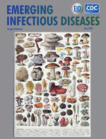
About the Cover
The Oldest Art
| EID | Breedlove B. The Oldest Art. Emerg Infect Dis. 2025;31(5):1064-1065. https://doi.org/10.3201/eid3105.ac3105 |
|---|---|
| AMA | Breedlove B. The Oldest Art. Emerging Infectious Diseases. 2025;31(5):1064-1065. doi:10.3201/eid3105.ac3105. |
| APA | Breedlove, B. (2025). The Oldest Art. Emerging Infectious Diseases, 31(5), 1064-1065. https://doi.org/10.3201/eid3105.ac3105. |


