Volume 30, Number 10—October 2024
Dispatch
Dengue Virus Serotype 3 Origins and Genetic Dynamics, Jamaica
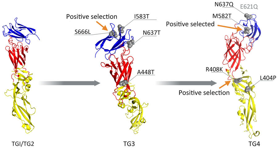
Figure 4

Figure 4. Envelope glycoprotein 3-dimensional structures (structure 7a3s; RCSB Protein Data Bank, https://www.rcsb.org) from dengue virus serotype 3 strains in Jamaica. Red indicates protein domain I, yellow indicates domain II, and blue indicates domain III. Gray spheres indicate mutations identified across various TGs. Arrows indicate mutations detected by site models. E621Q (faded text) is in the loop region not visible in the crystal structure. TG, temporal group.
1These first authors contributed equally to this article.
Page created: August 01, 2024
Page updated: September 23, 2024
Page reviewed: September 23, 2024
The conclusions, findings, and opinions expressed by authors contributing to this journal do not necessarily reflect the official position of the U.S. Department of Health and Human Services, the Public Health Service, the Centers for Disease Control and Prevention, or the authors' affiliated institutions. Use of trade names is for identification only and does not imply endorsement by any of the groups named above.