Volume 30, Number 9—September 2024
Synopsis
Morphologic and Molecular Identification of Human Ocular Infection Caused by Pelecitus Nematodes, Thailand
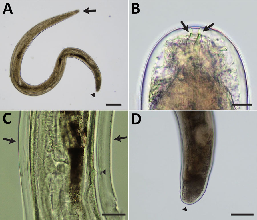
Figure 2

Figure 2. Light micrographs of Pelecitus sp. nematode isolated from the left eye of a 61-year-old man in Thailand. A) Curled female, 3.5 mm in length and 198 µm in width. Head (arrow) and tail (arrowhead) are indicated. Scale bar = 250 μm. B) Rounded anterior extremity with preesophageal cuticular ring (arrows). Scale bar = 10 μm. C) Postdeirid (arrowhead) at the posterior left side and lateral alae (arrows) at the posterior part. Scale bar = 50 μm. D) Rounded posterior extremity (arrowhead). Scale bar = 100 μm.
Page created: June 28, 2024
Page updated: August 20, 2024
Page reviewed: August 20, 2024
The conclusions, findings, and opinions expressed by authors contributing to this journal do not necessarily reflect the official position of the U.S. Department of Health and Human Services, the Public Health Service, the Centers for Disease Control and Prevention, or the authors' affiliated institutions. Use of trade names is for identification only and does not imply endorsement by any of the groups named above.