Disclaimer: Early release articles are not considered as final versions. Any changes will be reflected in the online version in the month the article is officially released.
Autochthonous Leishmania (Viannia) lainsoni in Dog, Rio de Janeiro State, Brazil, 2023
Isabela Cordeiro da Silva Santos, Daniel Moreira de Avelar, Luciana de Freitas Campos Miranda, Artur Augusto Velho Mendes Júnior, Lucas Keidel Oliveira, Luanna da Silva Ventura, Aline Fagundes da Silva, Fernanda Nunes Santos, Liliane de Fátima Antônio Oliveira, Rodrigo Caldas Menezes
1, and Andreza Pain Marcelino
1
Author affiliation: Author affiliations: Fundação Oswaldo Cruz, Rio de Janeiro, Brazil (I.C.d.S. Santos, L.d.F.C. Miranda, L.K. Oliveira, L.d.S. Ventura, A.F. da Silva, F.N. Santos, L.d.F.A. Oliveira, R.C. Menezes, A.P. Marcelino); Fundação Oswaldo Cruz, Belo Horizonte, Brazil (D.M. de Avelar); Instituto Carlos Chagas, Curitiba, Brazil (A.A.V.M. Júnior)
Main Article
Figure 1
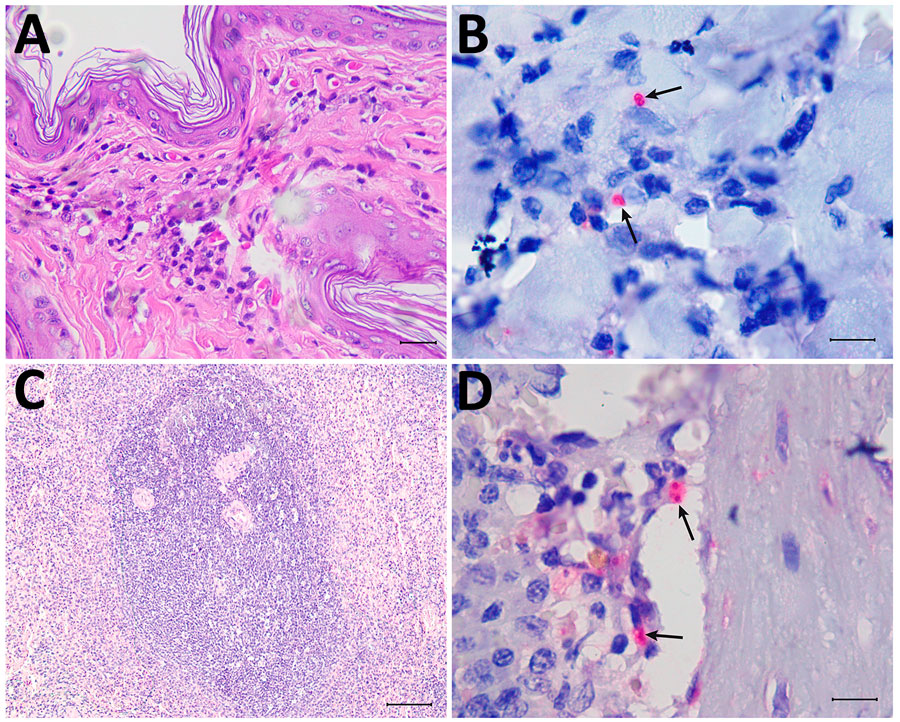
Figure 1. Histologic sections from autochthonous Leishmania (Viannia) lainsoni in dog (Canis familiaris), Rio de Janeiro state, Brazil, 2023. A, B) Skin of the examined dog: hyperkeratosis and moderate granulomatous infiltrate in the dermis are composed mainly of macrophages, with a smaller number of plasma cells and lymphocytes (A) and red-stained amastigotes in the cytoplasm of macrophages (arrows) (B). C, D) Spleen of the examined dog: lymphoid hyperplasia (C) and red-stained amastigotes in the cytoplasm of macrophages in the parenchyma (arrows) (D). A, C) Hematoxylin-eosin stain; B, D) immunohistochemistry. Scale bars indicate 10 µm.
Main Article
Page created: March 25, 2025
Page updated: April 15, 2025
Page reviewed: April 15, 2025
The conclusions, findings, and opinions expressed by authors contributing to this journal do not necessarily reflect the official position of the U.S. Department of Health and Human Services, the Public Health Service, the Centers for Disease Control and Prevention, or the authors' affiliated institutions. Use of trade names is for identification only and does not imply endorsement by any of the groups named above.