Disclaimer: Early release articles are not considered as final versions. Any changes will be reflected in the online version in the month the article is officially released.
Volume 31, Number 5—May 2025
Dispatch
Administration of L-type Bovine Spongiform Encephalopathy to Macaques to Evaluate Zoonotic Potential
Suggested citation for this article
Abstract
We administered L-type bovine spongiform encephalopathy prions to macaques to determine their potential for transmission to humans. After 75 months, no clinical symptoms appeared, and prions were undetectable in any tissue by Western blot or immunohistochemistry. Protein misfolding cyclic amplification, however, revealed prions in the nerve and lymphoid tissues.
Worldwide emergence of classical bovine spongiform encephalopathy (C-BSE) is associated with variant Creutzfeldt-Jakob disease in humans (1). Two other naturally occurring BSE variants have been identified, L-type (L-BSE) and H-type. Studies using transgenic mice expressing human normal prion protein (PrPC) (2) and primates (3–5) have demonstrated that L-BSE is more virulent than C-BSE. Although L-BSE is orally transmissible to minks (6), cattle (7), and mouse lemurs (5), transmissibility to cynomolgus macaques, a suitable model for investigating human susceptibility to prions, remains unclear. We orally inoculated cynomolgus macaques with L-BSE prions and explored the presence of abnormal prion proteins (PrPSc) in tissues using protein misfolding cyclic amplification (PMCA) along with Western blot (WB) and immunohistochemistry (IHC). PMCA markedly accelerates prion replication in vitro, and its products retain the biochemical properties and transmissibility of seed prion strains (8).
Two macaques orally inoculated with L-BSE prions remained asymptomatic and healthy but were euthanized and autopsied at 75 months postinoculation. WB showed no PrPSc accumulation in any tissue (Table), IHC revealed no PrPSc accumulation, hematoxylin and eosin staining revealed no spongiform changes in brain sections, and pathologic examination revealed no abnormalities.
We next attempted to detect PrPSc using PMCA, performed as previously described (9), with minor modifications (Appendix). First, we evaluated the sensitivity of PMCA. Using serial amplification with 10-fold stepwise dilutions of prion-infected brain homogenates as seeds, we amplified PrPSc-like proteinase K (PK)–resistant prion protein (PrPres) from a 10−7 dilution of 10% brain homogenate (BH) obtained from macaque intracerebrally inoculated with L-BSE prions in the fifth amplification round (Figure 1, panel A). This method also enabled propagation of PrPres from a 10−8 dilution of BH from C-BSE–affected cattle during the second amplification round (Figure 1, panel B), suggesting PMCA’s higher efficiency and sensitivity for detecting C-BSE prions than macaque L-BSE prions.
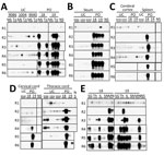
We attempted to detect prions in the lymphoid and nervous systems, among other tissues, of the 2 orally inoculated macaques using refined PMCA (Figure 2; Appendix Figure, Table). In lymphoid tissue samples prepared using sodium phosphotungstic acid precipitation (Appendix), we amplified PrPres in the inguinal and mesenteric lymph nodes, ileum, and tonsils of both macaques (Figure 2, panels A, B), as well as in the spleen of 1 macaque (#18) and the thymus of the other (#19), in the second or third amplification round of PMCA (Figure 2, panel C). We observed no PrPres in the submandibular lymph nodes (Appendix Figure 1). Examining the central nervous system, we observed no PrPres amplification in the cerebral cortex (Figure 2, panel C), whether seeded with phosphate-buffered saline homogenates or phosphotungstic acid precipitates. The spinal cord showed no PrPres amplification upon ethanol precipitation. However, PrPres was amplified in the cervical spinal cord of macaque #19 and in the thoracic spinal cord of both macaques with phosphate-buffered saline homogenates (Figure 2, panel D). We also confirmed PrPres in the median nerve of both macaques but not in the sciatic nerve (Figure 2, panel E). We noted PrPres signals in the submandibular glands of both animals. In contrast, we found no PrPres amplification in any tissues from uninoculated control macaques.
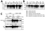
PrPres obtained from the orally inoculated macaques exhibited diverse banding patterns distinct from those generated by PMCA using L-BSE–affected cattle BH and L-BSE intracerebrally inoculated macaque BH as seeds (Figure 3, panels A–C). Of note, the lowest-molecular-weight PrPres variants from the ileum, spleen, inguinal lymph nodes, thoracic cord, submaxillary gland, and mesenteric lymph nodes of orally inoculated macaques exhibited remarkable PK resistance similarity and banding patterns indistinguishable from those of PrPres generated by PMCA with C-BSE–affected cattle BH as a seed (Figure 3, panel C, Appendix Figures 2 and 3). In contrast, the higher-molecular-weight PrPres variants from the ileum of macaque #18 exhibited a unique banding pattern distinct from those of L-BSE, C-BSE, and H-type BSE prions (Figure 3, panel C). Banding patterns and PK resistance of PrPres amplified from L-BSE–affected cattle BH and L-BSE intracerebrally inoculated macaque BH were notably similar.
This PMCA method was initially designed for the high-sensitivity detection of L-BSE intracerebrally inoculated macaque PrPSc but was even more efficient and sensitive in detecting bovine C-BSE PrPSc (Figure 1, panel B). Therefore, we believe that this method enabled the detection of both C-BSE–like PrPSc and potentially novel PrPSc variants.
We noted no detectable evidence of PrPSc by WB or IHC in any tissues of L-BSE orally inoculated macaques. Nevertheless, PMCA successfully amplified PrPres from lymphatic and neural tissues. The PrPres exhibited electrophoretic patterns distinct from those detected by PMCA using L-BSE–affected cattle BH as the seed (Figure 3, panel C), indicating that the PrPSc used as the template for PrPres amplification in orally inoculated macaques did not originate from the bovine L-BSE prions used as inoculum. Instead, PrPSc were newly generated by the conversion of macaque PrPC by bovine L-BSE prions. Our results provide strong evidence that L-BSE can infect macaques via the oral route.
We found no evidence that PrPSc reached the brain in orally inoculated macaques; however, the macaques euthanized 6 years postinoculation might have been in the preclinical period. At low infection levels, lymph nodes play a vital role in prion spread to the central nervous system (11). Therefore, had the macaques been maintained for a longer period, they might have developed prion disease. Retrospective surveillance studies using the appendix and tonsil tissues suggested a considerable number of humans harboring vCJD in a carrier state (12). Thus, we cannot exclude that L-BSE orally inoculated macaques could similarly remain in a potentially infectious state.
The brain of L-BSE intracerebrally inoculated macaque accumulated prions with biochemical properties resembling bovine L-BSE prions (Figure 3, panel C; Appendix Figure 2); however, we observed no PrPSc accumulation in lymphoid tissues by WB or IHC (4). In contrast, macaques orally inoculated with C-BSE prions showed PrPSc accumulation in lymphoid tissues, including the spleen, tonsils, and mesenteric lymph nodes by WB and IHC (13). In our study, L-BSE orally inoculated macaques harbored C-BSE–like prions in their lymphoid and neural tissues. Interspecies transmission of L-BSE prions to ovine PrP transgenic mice can result in a shift toward C-BSE–like properties (14,15). Our data suggest that L-BSE prions may alter biophysical and biochemical properties, depending on interspecies transmission and inoculation route, acquiring traits similar to those of C-BSE prions. This transformation might result from structural changes in the L-BSE prion to C-BSE–like prions and other lymphotropic prions within lymphoid tissues or from the selective propagation of low-level lymphotropic substrains within the L-BSE prion population.
The first limitation of our study is that the oral inoculation experiment involved only 2 macaques and tissues collected at 6 years postinoculation, before disease onset. Consequently, subsequent progression of prion disease symptoms remains speculative. A larger sample size and extended observation periods are required to conclusively establish infection in orally inoculated macaques. Furthermore, we performed no bioassays for PMCA-positive samples, leaving the relationship between PMCA results and infectious titers undefined. Considering that PrPres amplifications from tissues from the orally inoculated macaque tissues required 2 rounds of PMCA, the PrPSc levels in positive tissues might have been extremely low and undetectable in the bioassay.
Previous studies have demonstrated that L-BSE can be orally transmitted to cattle (7) and might have caused prion disease in farm-raised minks (6), indicating that L-BSE could naturally affect various animal species. Our findings suggest that L-BSE can also be orally transmitted to macaques. Therefore, current control measures aimed at preventing primary C-BSE in cattle and humans may also need to consider the potential risk of spontaneous L-BSE transmission.
Dr. Imamura is an associate professor in the Division of Microbiology, Department of Infectious Diseases, Faculty of Medicine, University of Miyazaki, Miyazaki, Miyazaki, Japan. His research interests are focused on elucidating the mechanisms underlying prion formation.
Acknowledgment
This study was supported by the Health Labor Sciences Research Grant (H29-Shokuhin-Ippan-004, 20KA1003, and 23KA1004).
References
- Will RG, Ironside JW, Zeidler M, Cousens SN, Estibeiro K, Alperovitch A, et al. A new variant of Creutzfeldt-Jakob disease in the UK. Lancet. 1996;347:921–5. DOIPubMedGoogle Scholar
- Béringue V, Herzog L, Reine F, Le Dur A, Casalone C, Vilotte JL, et al. Transmission of atypical bovine prions to mice transgenic for human prion protein. Emerg Infect Dis. 2008;14:1898–901. DOIPubMedGoogle Scholar
- Comoy EE, Casalone C, Lescoutra-Etchegaray N, Zanusso G, Freire S, Marcé D, et al. Atypical BSE (BASE) transmitted from asymptomatic aging cattle to a primate. PLoS One. 2008;3:
e3017 . DOIPubMedGoogle Scholar - Ono F, Tase N, Kurosawa A, Hiyaoka A, Ohyama A, Tezuka Y, et al. Atypical L-type bovine spongiform encephalopathy (L-BSE) transmission to cynomolgus macaques, a non-human primate. Jpn J Infect Dis. 2011;64:81–4. DOIPubMedGoogle Scholar
- Mestre-Francés N, Nicot S, Rouland S, Biacabe AG, Quadrio I, Perret-Liaudet A, et al. Oral transmission of L-type bovine spongiform encephalopathy in primate model. Emerg Infect Dis. 2012;18:142–5. DOIPubMedGoogle Scholar
- Baron T, Bencsik A, Biacabe AG, Morignat E, Bessen RA. Phenotypic similarity of transmissible mink encephalopathy in cattle and L-type bovine spongiform encephalopathy in a mouse model. Emerg Infect Dis. 2007;13:1887–94. DOIPubMedGoogle Scholar
- Okada H, Iwamaru Y, Imamura M, Miyazawa K, Matsuura Y, Masujin K, et al. Oral transmission of L-type bovine spongiform encephalopathy agent among cattle. Emerg Infect Dis. 2017;23:284–7. DOIPubMedGoogle Scholar
- Castilla J, Morales R, Saá P, Barria M, Gambetti P, Soto C. Cell-free propagation of prion strains. EMBO J. 2008;27:2557–66. DOIPubMedGoogle Scholar
- Murayama Y, Ono F, Shimozaki N, Shibata H. L-Arginine ethylester enhances in vitro amplification of PrP(Sc) in macaques with atypical L-type bovine spongiform encephalopathy and enables presymptomatic detection of PrP(Sc) in the bodily fluids. Biochem Biophys Res Commun. 2016;470:563–8. DOIPubMedGoogle Scholar
- Murayama Y, Yoshioka M, Masujin K, Okada H, Iwamaru Y, Imamura M, et al. Sulfated dextrans enhance in vitro amplification of bovine spongiform encephalopathy PrP(Sc) and enable ultrasensitive detection of bovine PrP(Sc). PLoS One. 2010;5:
e13152 . DOIPubMedGoogle Scholar - Kratzel C, Krüger D, Beekes M. Relevance of the regional lymph node in scrapie pathogenesis after peripheral infection of hamsters. BMC Vet Res. 2007;3:22. DOIPubMedGoogle Scholar
- Hilton DA, Ghani AC, Conyers L, Edwards P, McCardle L, Ritchie D, et al. Prevalence of lymphoreticular prion protein accumulation in UK tissue samples. J Pathol. 2004;203:733–9. DOIPubMedGoogle Scholar
- Herzog C, Rivière J, Lescoutra-Etchegaray N, Charbonnier A, Leblanc V, Salès N, et al. PrPTSE distribution in a primate model of variant, sporadic, and iatrogenic Creutzfeldt-Jakob disease. J Virol. 2005;79:14339–45. DOIPubMedGoogle Scholar
- Béringue V, Andréoletti O, Le Dur A, Essalmani R, Vilotte JL, Lacroux C, et al. A bovine prion acquires an epidemic bovine spongiform encephalopathy strain-like phenotype on interspecies transmission. J Neurosci. 2007;27:6965–71. DOIPubMedGoogle Scholar
- Baron T, Bencsik A, Morignat E. Prions of ruminants show distinct splenotropisms in an ovine transgenic mouse model. PLoS One. 2010;5:
e10310 . DOIPubMedGoogle Scholar
Figures
Table
Suggested citation for this article: Imamura M, Hagiwara K, Tobiume M, Ohno M, Iguchi H, Takatsuki H, et al. Administration of L-type bovine spongiform encephalopathy to macaques to evaluate zoonotic potential. Emerg Infect Dis. 2025 May [date cited]. https://doi.org/10.3201/eid3105.241257
Original Publication Date: April 15, 2025
1These authors were co–principal investigators.
Table of Contents – Volume 31, Number 5—May 2025
| EID Search Options |
|---|
|
|
|
|
|
|


Please use the form below to submit correspondence to the authors or contact them at the following address:
Morikazu Imamura, Division of Microbiology, Department of Infectious Diseases, Faculty of Medicine, University of Miyazaki, 5200 Kihara, Kiyotake, Miyazaki 889-1692, Japan
Top