Volume 9, Number 7—July 2003
Research
West Nile Virus in Farmed Alligators
Debra L. Miller* , Michael J. Mauel*, Charles Baldwin*, Gary Burtle*, Dallas Ingram*, Murray E. Hines*, and Kendal S. Frazier*
Figure 1
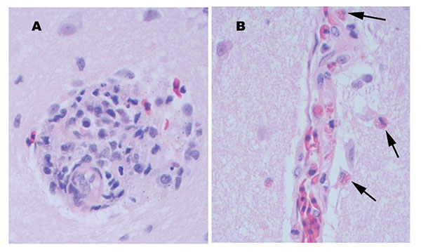
Figure 1. Perivascular changes observed within the brain of alligators infected with West Nile virus (400x). A. Perivascular infiltrates were composed of primarily lymphocytes, plasma cells, and macrophages in the hatchling alligator. B. Perivascular infiltrates were composed of primarily heterophils (arrows) in juvenile alligators.
Page created: December 22, 2010
Page updated: December 22, 2010
Page reviewed: December 22, 2010
The conclusions, findings, and opinions expressed by authors contributing to this journal do not necessarily reflect the official position of the U.S. Department of Health and Human Services, the Public Health Service, the Centers for Disease Control and Prevention, or the authors' affiliated institutions. Use of trade names is for identification only and does not imply endorsement by any of the groups named above.