Perspective
Emerging Trends in International Law Concerning Global Infectious Disease Control
International cooperation has become critical in controlling infectious diseases. In this article, I examine emerging trends in international law concerning global infectious disease control. The role of international law in horizontal and vertical governance responses to infectious disease control is conceptualized; the historical development of international law regarding infectious diseases is described; and important shifts in how states, international institutions, and nonstate organizations use international law in the context of infectious disease control today are analyzed. The growing importance of international trade law and the development of global governance mechanisms, most prominently in connection with increasing access to drugs and other medicines in unindustrialized countries, are emphasized. Traditional international legal approaches to infectious disease control—embodied in the International Health Regulations—may be moribund.
| EID | Fidler DP. Emerging Trends in International Law Concerning Global Infectious Disease Control. Emerg Infect Dis. 2003;9(3):285-290. https://doi.org/10.3201/eid0903.020336 |
|---|---|
| AMA | Fidler DP. Emerging Trends in International Law Concerning Global Infectious Disease Control. Emerging Infectious Diseases. 2003;9(3):285-290. doi:10.3201/eid0903.020336. |
| APA | Fidler, D. P. (2003). Emerging Trends in International Law Concerning Global Infectious Disease Control. Emerging Infectious Diseases, 9(3), 285-290. https://doi.org/10.3201/eid0903.020336. |
Synopses
Enterovirus 71 Outbreaks, Taiwan: Occurrence and Recognition
| EID | Lin T, Twu S, Ho M, Chang L, Lee C. Enterovirus 71 Outbreaks, Taiwan: Occurrence and Recognition. Emerg Infect Dis. 2003;9(3):291-293. https://doi.org/10.3201/eid0903.020285 |
|---|---|
| AMA | Lin T, Twu S, Ho M, et al. Enterovirus 71 Outbreaks, Taiwan: Occurrence and Recognition. Emerging Infectious Diseases. 2003;9(3):291-293. doi:10.3201/eid0903.020285. |
| APA | Lin, T., Twu, S., Ho, M., Chang, L., & Lee, C. (2003). Enterovirus 71 Outbreaks, Taiwan: Occurrence and Recognition. Emerging Infectious Diseases, 9(3), 291-293. https://doi.org/10.3201/eid0903.020285. |
Electron Microscopy for Rapid Diagnosis of Emerging Infectious Agents
Diagnostic electron microscopy has two advantages over enzyme-linked immunosorbent assay and nucleic acid amplification tests. After a simple and fast negative stain preparation, the undirected, “open view” of electron microscopy allows rapid morphologic identification and differential diagnosis of different agents contained in the specimen. Details for efficient sample collection, preparation, and particle enrichment are given. Applications of diagnostic electron microscopy in clinically or epidemiologically critical situations as well as in bioterrorist events are discussed. Electron microscopy can be applied to many body samples and can also hasten routine cell culture diagnosis. To exploit the potential of diagnostic electron microscopy fully, it should be quality controlled, applied as a frontline method, and be coordinated and run in parallel with other diagnostic techniques.
| EID | Hazelton PR, Gelderblom HR. Electron Microscopy for Rapid Diagnosis of Emerging Infectious Agents. Emerg Infect Dis. 2003;9(3):294-303. https://doi.org/10.3201/eid0903.020327 |
|---|---|
| AMA | Hazelton PR, Gelderblom HR. Electron Microscopy for Rapid Diagnosis of Emerging Infectious Agents. Emerging Infectious Diseases. 2003;9(3):294-303. doi:10.3201/eid0903.020327. |
| APA | Hazelton, P. R., & Gelderblom, H. R. (2003). Electron Microscopy for Rapid Diagnosis of Emerging Infectious Agents. Emerging Infectious Diseases, 9(3), 294-303. https://doi.org/10.3201/eid0903.020327. |
Research
Influenza AH1N2 Viruses, United Kingdom, 2001–02 Influenza Season
During the winter of 2001–02, influenza AH1N2 viruses were detected for the first time in humans in the U.K. The H1N2 viruses co-circulated with H3N2 viruses and a very small number of H1N1 viruses and were isolated in the community and hospitalized patients, predominantly from children <15 years of age. Characterization of H1N2 viruses indicated that they were antigenically and genetically homogeneous, deriving the hemagglutinin (HA) gene from recently circulating A/New Caledonia/20/99-like H1N1 viruses, whereas the other seven genes originated from recently circulating H3N2 viruses. Retrospective reverse transcription-polymerase chain reaction analysis of influenza A H1 viruses isolated in the U.K. during the previous winter identified a single H1N2 virus, isolated in March 2001, indicating that H1N2 viruses did not widely circulate in the U.K. before September 2001. The reassortment event is estimated to have occurred between 1999 and early 2001, and the emergence of H1N2 viruses in humans reinforces the need for frequent surveillance of circulating viruses.
| EID | Ellis JS, Alvarez-Aguero A, Gregory V, Lin YP, Hay A, Zambon MC. Influenza AH1N2 Viruses, United Kingdom, 2001–02 Influenza Season. Emerg Infect Dis. 2003;9(3):304-310. https://doi.org/10.3201/eid0903.020404 |
|---|---|
| AMA | Ellis JS, Alvarez-Aguero A, Gregory V, et al. Influenza AH1N2 Viruses, United Kingdom, 2001–02 Influenza Season. Emerging Infectious Diseases. 2003;9(3):304-310. doi:10.3201/eid0903.020404. |
| APA | Ellis, J. S., Alvarez-Aguero, A., Gregory, V., Lin, Y. P., Hay, A., & Zambon, M. C. (2003). Influenza AH1N2 Viruses, United Kingdom, 2001–02 Influenza Season. Emerging Infectious Diseases, 9(3), 304-310. https://doi.org/10.3201/eid0903.020404. |
Experimental Infection of North American Birds with the New York 1999 Strain of West Nile Virus
To evaluate transmission dynamics, we exposed 25 bird species to West Nile virus (WNV) by infectious mosquito bite. We monitored viremia titers, clinical outcome, WNV shedding (cloacal and oral), seroconversion, virus persistence in organs, and susceptibility to oral and contact transmission. Passeriform and charadriiform birds were more reservoir competent (a derivation of viremia data) than other species tested. The five most competent species were passerines: Blue Jay (Cyanocitta cristata), Common Grackle (Quiscalus quiscula), House Finch (Carpodacus mexicanus), American Crow (Corvus brachyrhynchos), and House Sparrow (Passer domesticus). Death occurred in eight species. Cloacal shedding of WNV was observed in 17 of 24 species, and oral shedding in 12 of 14 species. We observed contact transmission among four species and oral in five species. Persistent WNV infections were found in tissues of 16 surviving birds. Our observations shed light on transmission ecology of WNV and will benefit surveillance and control programs.
| EID | Komar N, Langevin S, Hinten S, Nemeth NM, Edwards E, Hettler DL, et al. Experimental Infection of North American Birds with the New York 1999 Strain of West Nile Virus. Emerg Infect Dis. 2003;9(3):311-322. https://doi.org/10.3201/eid0903.020628 |
|---|---|
| AMA | Komar N, Langevin S, Hinten S, et al. Experimental Infection of North American Birds with the New York 1999 Strain of West Nile Virus. Emerging Infectious Diseases. 2003;9(3):311-322. doi:10.3201/eid0903.020628. |
| APA | Komar, N., Langevin, S., Hinten, S., Nemeth, N. M., Edwards, E., Hettler, D. L....Bunning, M. L. (2003). Experimental Infection of North American Birds with the New York 1999 Strain of West Nile Virus. Emerging Infectious Diseases, 9(3), 311-322. https://doi.org/10.3201/eid0903.020628. |
Emergence of Ceftriaxone-Resistant Salmonella Isolates and Rapid Spread of Plasmid-Encoded CMY-2–Like Cephalosporinase, Taiwan
Of 384 Salmonella isolates collected from 1997 to 2000 in a university hospital in Taiwan, six ceftriaxone-resistant isolates of Salmonella enterica serovar Typhimurium were found in two patients in 2000. The resistance determinants were on conjugative plasmids that encoded a CMY-2–like cephalosporinase. During the study period, the proportion of CMY-2–like enzyme producers among Escherichia coli increased rapidly from 0.2% in early 1999 to >4.0% in late 2000. Klebsiella pneumoniae isolates producing a CMY-2–like β-lactamase did not emerge until 2000. The presence of blaCMY-containing plasmids with an identical restriction pattern from Salmonella, E. coli, and K. pneumoniae isolates was found, which suggests interspecies spread and horizontal transfer of the resistance determinant. Various nosocomial and community-acquired infections were associated with the CMY-2–like enzyme producers. Our study suggests that the spread of plasmid-mediated CMY-2–like β-lactamases is an emerging threat to hospitalized patients and the public in Taiwan.
| EID | Yan J, Ko W, Chiu C, Tsai S, Wu H, Wu J. Emergence of Ceftriaxone-Resistant Salmonella Isolates and Rapid Spread of Plasmid-Encoded CMY-2–Like Cephalosporinase, Taiwan. Emerg Infect Dis. 2003;9(3):323-328. https://doi.org/10.3201/eid0903.010410 |
|---|---|
| AMA | Yan J, Ko W, Chiu C, et al. Emergence of Ceftriaxone-Resistant Salmonella Isolates and Rapid Spread of Plasmid-Encoded CMY-2–Like Cephalosporinase, Taiwan. Emerging Infectious Diseases. 2003;9(3):323-328. doi:10.3201/eid0903.010410. |
| APA | Yan, J., Ko, W., Chiu, C., Tsai, S., Wu, H., & Wu, J. (2003). Emergence of Ceftriaxone-Resistant Salmonella Isolates and Rapid Spread of Plasmid-Encoded CMY-2–Like Cephalosporinase, Taiwan. Emerging Infectious Diseases, 9(3), 323-328. https://doi.org/10.3201/eid0903.010410. |
Bartonella henselae in Ixodes ricinus Ticks (Acari: Ixodida) Removed from Humans, Belluno Province, Italy
The potential role of ticks as vectors of Bartonella species has recently been suggested. In this study, we investigated the presence of Bartonella species in 271 ticks removed from humans in Belluno Province, Italy. By using primers derived from the 60-kDa heat shock protein gene sequences, Bartonella DNA was amplified and sequenced from four Ixodes ricinus ticks (1.48%). To confirm this finding, we performed amplification and partial sequencing of the pap31 protein and the cell division protein FtsZ encoding genes. This process allowed us to definitively identify B. henselae (genotype Houston-1) DNA in the four ticks. Detection of B. henselae in these ticks might represent a highly sensitive form of xenodiagnosis. B. henselae is the first human-infecting Bartonella identified from Ixodes ricinus, a common European tick and the vector of various tickborne pathogens. The role of ticks in the transmission of bartonellosis should be further investigated.
| EID | Sanogo YO, Zeaiter Z, Caruso G, Merola F, Shpynov S, Brouqui P, et al. Bartonella henselae in Ixodes ricinus Ticks (Acari: Ixodida) Removed from Humans, Belluno Province, Italy. Emerg Infect Dis. 2003;9(3):329-332. https://doi.org/10.3201/eid0903.020133 |
|---|---|
| AMA | Sanogo YO, Zeaiter Z, Caruso G, et al. Bartonella henselae in Ixodes ricinus Ticks (Acari: Ixodida) Removed from Humans, Belluno Province, Italy. Emerging Infectious Diseases. 2003;9(3):329-332. doi:10.3201/eid0903.020133. |
| APA | Sanogo, Y. O., Zeaiter, Z., Caruso, G., Merola, F., Shpynov, S., Brouqui, P....Raoult, D. (2003). Bartonella henselae in Ixodes ricinus Ticks (Acari: Ixodida) Removed from Humans, Belluno Province, Italy. Emerging Infectious Diseases, 9(3), 329-332. https://doi.org/10.3201/eid0903.020133. |
New Lyssavirus Genotype from the Lesser Mouse-eared Bat (Myotis blythi), Kyrghyzstan
The Aravan virus was isolated from a Lesser Mouse-eared Bat (Myotis blythi) in the Osh region of Kyrghyzstan, central Asia, in 1991. We determined the complete sequence of the nucleoprotein (N) gene and compared it with those of 26 representative lyssaviruses obtained from databases. The Aravan virus was distinguished from seven distinct genotypes on the basis of nucleotide and amino acid identity. Phylogenetic analysis based on both nucleotide and amino acid sequences showed that the Aravan virus was more closely related to genotypes 4, 5, and—to a lesser extent—6, which circulates among insectivorus bats in Europe and Africa. The Aravan virus does not belong to any of the seven known genotypes of lyssaviruses, namely, rabies, Lagos bat, Mokola, and Duvenhage viruses and European bat lyssavirus 1, European bat lyssavirus 2, and Australian bat lyssavirus. Based on these data, we propose a new genotype for the Lyssavirus genus.
| EID | Arai YT, Kuzmin IV, Kameoka Y, Botvinkin AD. New Lyssavirus Genotype from the Lesser Mouse-eared Bat (Myotis blythi), Kyrghyzstan. Emerg Infect Dis. 2003;9(3):333-337. https://doi.org/10.3201/eid0903.020252 |
|---|---|
| AMA | Arai YT, Kuzmin IV, Kameoka Y, et al. New Lyssavirus Genotype from the Lesser Mouse-eared Bat (Myotis blythi), Kyrghyzstan. Emerging Infectious Diseases. 2003;9(3):333-337. doi:10.3201/eid0903.020252. |
| APA | Arai, Y. T., Kuzmin, I. V., Kameoka, Y., & Botvinkin, A. D. (2003). New Lyssavirus Genotype from the Lesser Mouse-eared Bat (Myotis blythi), Kyrghyzstan. Emerging Infectious Diseases, 9(3), 333-337. https://doi.org/10.3201/eid0903.020252. |
Molecular Detection of Bartonella quintana, B. koehlerae, B. henselae, B. clarridgeiae, Rickettsia felis, and Wolbachia pipientis in Cat Fleas, France
| EID | Rolain J, Franc M, Davoust B, Raoult D. Molecular Detection of Bartonella quintana, B. koehlerae, B. henselae, B. clarridgeiae, Rickettsia felis, and Wolbachia pipientis in Cat Fleas, France. Emerg Infect Dis. 2003;9(3):339-342. https://doi.org/10.3201/eid0903.020278 |
|---|---|
| AMA | Rolain J, Franc M, Davoust B, et al. Molecular Detection of Bartonella quintana, B. koehlerae, B. henselae, B. clarridgeiae, Rickettsia felis, and Wolbachia pipientis in Cat Fleas, France. Emerging Infectious Diseases. 2003;9(3):339-342. doi:10.3201/eid0903.020278. |
| APA | Rolain, J., Franc, M., Davoust, B., & Raoult, D. (2003). Molecular Detection of Bartonella quintana, B. koehlerae, B. henselae, B. clarridgeiae, Rickettsia felis, and Wolbachia pipientis in Cat Fleas, France. Emerging Infectious Diseases, 9(3), 339-342. https://doi.org/10.3201/eid0903.020278. |
European Echinococcosis Registry: Human Alveolar Echinococcosis, Europe, 1982–2000
Surveillance for alveolar echinococcosis in central Europe was initiated in 1998. On a voluntary basis, 559 patients were reported to the registry. Most cases originated from rural communities in regions from eastern France to western Austria; single cases were reported far away from the disease-“endemic” zone throughout central Europe. Of 210 patients, 61.4% were involved in vocational or part-time farming, gardening, forestry, or hunting. Patients were diagnosed at a mean age of 52.5 years; 78% had symptoms. Alveolar echinococcosis primarily manifested as a liver disease. Of the 559 patients, 190 (34%) were already affected by spread of the parasitic larval tissue. Of 408 (73%) patients alive in 2000, 4.9% were cured. The increasing prevalence of Echinococcus multilocularis in foxes in rural and urban areas of central Europe and the occurrence of cases outside the alveolar echinococcosis–endemic regions suggest that this disease deserves increased attention.
| EID | Kern P, Bardonnet K, Renner E, Auer H, Pawlowski Z, Ammann RW, et al. European Echinococcosis Registry: Human Alveolar Echinococcosis, Europe, 1982–2000. Emerg Infect Dis. 2003;9(3):343-349. https://doi.org/10.3201/eid0903.020341 |
|---|---|
| AMA | Kern P, Bardonnet K, Renner E, et al. European Echinococcosis Registry: Human Alveolar Echinococcosis, Europe, 1982–2000. Emerging Infectious Diseases. 2003;9(3):343-349. doi:10.3201/eid0903.020341. |
| APA | Kern, P., Bardonnet, K., Renner, E., Auer, H., Pawlowski, Z., Ammann, R. W....Kern, P. (2003). European Echinococcosis Registry: Human Alveolar Echinococcosis, Europe, 1982–2000. Emerging Infectious Diseases, 9(3), 343-349. https://doi.org/10.3201/eid0903.020341. |
Tularemia on Martha’s Vineyard: Seroprevalence and Occupational Risk
We conducted a serosurvey of landscapers to determine if they were at increased risk for exposure to Francisella tularensis and to determine risk factors for infection. In Martha’s Vineyard, Massachusetts, landscapers (n=132) were tested for anti–F. tularensis antibody and completed a questionnaire. For comparison, serum samples from three groups of nonlandscaper Martha’s Vineyard residents (n=103, 99, and 108) were tested. Twelve landscapers (9.1%) were seropositive, compared with one person total from the comparison groups (prevalence ratio 9.0; 95% confidence interval 1.2 to 68.1; p=0.02). Of landscapers who used a power blower, 15% were seropositive, compared to 2% who did not use a power blower (prevalence ratio 9.2; 95% confidence interval 1.2 to 69.0; p=0.02). Seropositive landscapers worked more hours per week mowing and weed-whacking and mowed more lawns per week than their seronegative counterparts. Health-care workers in tularemia-endemic areas should consider tularemia as a diagnosis for landscapers with a febrile illness.
| EID | Feldman KA, Stiles-Enos D, Julian K, Matyas BT, Telford SR, Chu MC, et al. Tularemia on Martha’s Vineyard: Seroprevalence and Occupational Risk. Emerg Infect Dis. 2003;9(3):350-354. https://doi.org/10.3201/eid0903.020462 |
|---|---|
| AMA | Feldman KA, Stiles-Enos D, Julian K, et al. Tularemia on Martha’s Vineyard: Seroprevalence and Occupational Risk. Emerging Infectious Diseases. 2003;9(3):350-354. doi:10.3201/eid0903.020462. |
| APA | Feldman, K. A., Stiles-Enos, D., Julian, K., Matyas, B. T., Telford, S. R., Chu, M. C....Hayes, E. B. (2003). Tularemia on Martha’s Vineyard: Seroprevalence and Occupational Risk. Emerging Infectious Diseases, 9(3), 350-354. https://doi.org/10.3201/eid0903.020462. |
Epidemiology of Meningococcal Disease, New York City, 1989–2000
Study of the epidemiologic trends in meningococcal disease is important in understanding infection dynamics and developing timely and appropriate public health interventions. We studied surveillance data from the New York City Department of Health and Mental Hygiene, which showed that during 1989–2000 a decrease occurred in both the proportion of patients with serogroup B infection (from 28% to 13% of reported cases; p<0.01) and the rate of serogroup B infection (from 0.25/100,000 to 0.08/100,000; p<0.01). We also noted an increased proportion (from 3% to 39%; p<0.01) and rate of serogroup Y infection (from 0.02/100,000 to 0.23/100,000; p<0.01). Median patient age increased (from 15 to 30 years; p<0.01). The case-fatality rate for the period was 17%. As more effective meningococcal vaccines become available, recommendations for their use in nonepidemic settings should consider current epidemiologic trends, particularly changes in age and serogroup distribution of meningococcal infections.
| EID | Moura AS, Pablos-Méndez A, Layton M, Weiss D. Epidemiology of Meningococcal Disease, New York City, 1989–2000. Emerg Infect Dis. 2003;9(3):355-361. https://doi.org/10.3201/eid0903.020071 |
|---|---|
| AMA | Moura AS, Pablos-Méndez A, Layton M, et al. Epidemiology of Meningococcal Disease, New York City, 1989–2000. Emerging Infectious Diseases. 2003;9(3):355-361. doi:10.3201/eid0903.020071. |
| APA | Moura, A. S., Pablos-Méndez, A., Layton, M., & Weiss, D. (2003). Epidemiology of Meningococcal Disease, New York City, 1989–2000. Emerging Infectious Diseases, 9(3), 355-361. https://doi.org/10.3201/eid0903.020071. |
Amplification of the Sylvatic Cycle of Dengue Virus Type 2, Senegal, 1999–2000: Entomologic Findings and Epidemiologic Considerations
After 8 years of silence, dengue virus serotype 2 (DENV-2) reemerged in southeastern Senegal in 1999. Sixty-four DENV-2 strains were isolated in 1999 and 9 strains in 2000 from mosquitoes captured in the forest gallery and surrounding villages. Isolates were obtained from previously described vectors, Aedes furcifer, Ae. taylori, Ae. luteocephalus, and—for the first time in Senegal—from Ae. aegypti and Ae. vittatus. A retrospective analysis of sylvatic DENV-2 outbreaks in Senegal during the last 28 years of entomologic investigations shows that amplifications are periodic, with intervening, silent intervals of 5–8 years. No correlation was found between sylvatic DENV-2 emergence and rainfall amount. For sylvatic DENV-2 vectors, rainfall seems to particularly affect virus amplification that occurs at the end of the rainy season, from October to November. Data obtained from investigation of preimaginal (i.e., nonadult) mosquitoes suggest a secondary transmission cycle involving mosquitoes other than those identified previously as vectors.
| EID | Diallo M, Ba Y, Sall AA, Diop OM, Ndione JA, Mondo M, et al. Amplification of the Sylvatic Cycle of Dengue Virus Type 2, Senegal, 1999–2000: Entomologic Findings and Epidemiologic Considerations. Emerg Infect Dis. 2003;9(3):362-367. https://doi.org/10.3201/eid0903.020219 |
|---|---|
| AMA | Diallo M, Ba Y, Sall AA, et al. Amplification of the Sylvatic Cycle of Dengue Virus Type 2, Senegal, 1999–2000: Entomologic Findings and Epidemiologic Considerations. Emerging Infectious Diseases. 2003;9(3):362-367. doi:10.3201/eid0903.020219. |
| APA | Diallo, M., Ba, Y., Sall, A. A., Diop, O. M., Ndione, J. A., Mondo, M....Mathiot, C. (2003). Amplification of the Sylvatic Cycle of Dengue Virus Type 2, Senegal, 1999–2000: Entomologic Findings and Epidemiologic Considerations. Emerging Infectious Diseases, 9(3), 362-367. https://doi.org/10.3201/eid0903.020219. |
Dispatches
Rabies in Sri Lanka: Splendid Isolation
Rabies virus exists in dogs on Sri Lanka as a single, minimally divergent lineage only distantly related to other rabies virus lineages in Asia. Stable, geographically isolated virus populations are susceptible to local extinction. A fully implemented rabies-control campaign could make Sri Lanka the first Asian country in >30 years to become free of rabies virus.
| EID | Nanayakkara S, Smith JS, Rupprecht CE. Rabies in Sri Lanka: Splendid Isolation. Emerg Infect Dis. 2003;9(3):368-371. https://doi.org/10.3201/eid0903.020545 |
|---|---|
| AMA | Nanayakkara S, Smith JS, Rupprecht CE. Rabies in Sri Lanka: Splendid Isolation. Emerging Infectious Diseases. 2003;9(3):368-371. doi:10.3201/eid0903.020545. |
| APA | Nanayakkara, S., Smith, J. S., & Rupprecht, C. E. (2003). Rabies in Sri Lanka: Splendid Isolation. Emerging Infectious Diseases, 9(3), 368-371. https://doi.org/10.3201/eid0903.020545. |
Human Metapneumovirus in Severe Respiratory Syncytial Virus Bronchiolitis
Reverse transcription-polymerase chain reaction was used to detect segments of the M (matrix), N (nucleoprotein), and F (fusion) genes of human metapneumovirus in bronchoalveolar fluid from 30 infants with severe respiratory syncytial virus bronchiolitis. Seventy percent of them were coinfected with metapneumovirus. Such coinfection might be a factor influencing the severity of bronchiolitis.
| EID | Greensill J, McNamara PS, Dove W, Flanagan B, Smyth RL, Hart CA. Human Metapneumovirus in Severe Respiratory Syncytial Virus Bronchiolitis. Emerg Infect Dis. 2003;9(3):372-375. https://doi.org/10.3201/eid0903.020289 |
|---|---|
| AMA | Greensill J, McNamara PS, Dove W, et al. Human Metapneumovirus in Severe Respiratory Syncytial Virus Bronchiolitis. Emerging Infectious Diseases. 2003;9(3):372-375. doi:10.3201/eid0903.020289. |
| APA | Greensill, J., McNamara, P. S., Dove, W., Flanagan, B., Smyth, R. L., & Hart, C. A. (2003). Human Metapneumovirus in Severe Respiratory Syncytial Virus Bronchiolitis. Emerging Infectious Diseases, 9(3), 372-375. https://doi.org/10.3201/eid0903.020289. |
Persistence of Virus-Reactive Serum Immunoglobulin M Antibody in Confirmed West Nile Virus Encephalitis Cases
Twenty-nine laboratory-confirmed West Nile virus (WNV) encephalitis patients were bled serially so that WNV-reactive immunoglobulin (Ig) M activity could be determined. Of those patients bled, 7 (60%) of 12 had anti-WNV IgM at approximately 500 days after onset. Clinicians should be cautious when interpreting serologic results from early season WNV IgM-positive patients.
| EID | Roehrig JT, Nash D, Maldin B, Labowitz A, Martin DA, Lanciotti RS, et al. Persistence of Virus-Reactive Serum Immunoglobulin M Antibody in Confirmed West Nile Virus Encephalitis Cases. Emerg Infect Dis. 2003;9(3):376-379. https://doi.org/10.3201/eid0903.020531 |
|---|---|
| AMA | Roehrig JT, Nash D, Maldin B, et al. Persistence of Virus-Reactive Serum Immunoglobulin M Antibody in Confirmed West Nile Virus Encephalitis Cases. Emerging Infectious Diseases. 2003;9(3):376-379. doi:10.3201/eid0903.020531. |
| APA | Roehrig, J. T., Nash, D., Maldin, B., Labowitz, A., Martin, D. A., Lanciotti, R. S....Campbell, G. L. (2003). Persistence of Virus-Reactive Serum Immunoglobulin M Antibody in Confirmed West Nile Virus Encephalitis Cases. Emerging Infectious Diseases, 9(3), 376-379. https://doi.org/10.3201/eid0903.020531. |
Isolation of Escherichia coli O157:H7 from Intact Colon Fecal Samples of Swine
Escherichia coli O157:H7 was recovered from colon fecal samples of pigs. Polymerase chain reaction confirmed two genotypes: isolates harboring the eaeA, stx1, and stx2 genes and isolates harboring the eaeA, stx1, and hly933 genes. We demonstrate that swine in the United States can harbor potentially pathogenic E. coli O157:H7.
| EID | Feder I, Wallace FM, Gray JT, Fratamico P, Fedorka-Cray PJ, Pearce RA, et al. Isolation of Escherichia coli O157:H7 from Intact Colon Fecal Samples of Swine. Emerg Infect Dis. 2003;9(3):380-383. https://doi.org/10.3201/eid0903.020350 |
|---|---|
| AMA | Feder I, Wallace FM, Gray JT, et al. Isolation of Escherichia coli O157:H7 from Intact Colon Fecal Samples of Swine. Emerging Infectious Diseases. 2003;9(3):380-383. doi:10.3201/eid0903.020350. |
| APA | Feder, I., Wallace, F. M., Gray, J. T., Fratamico, P., Fedorka-Cray, P. J., Pearce, R. A....Luchansky, J. B. (2003). Isolation of Escherichia coli O157:H7 from Intact Colon Fecal Samples of Swine. Emerging Infectious Diseases, 9(3), 380-383. https://doi.org/10.3201/eid0903.020350. |
Echinococcus multilocularis: An Emerging Pathogen in Hungary and Central Eastern Europe?
Echinococcus multilocularis, the causative agent of human alveolar echinococcosis, is reported for the first time in Red Foxes (Vulpes vulpes) in Hungary. This parasite may be spreading eastward because the population of foxes has increased because of human interventions, and this spread may result in the emergence of alveolar echinococcosis in Central Eastern Europe.
| EID | Sréter T, Széll Z, Egyed Z, Varga I. Echinococcus multilocularis: An Emerging Pathogen in Hungary and Central Eastern Europe?. Emerg Infect Dis. 2003;9(3):384-386. https://doi.org/10.3201/eid0903.020320 |
|---|---|
| AMA | Sréter T, Széll Z, Egyed Z, et al. Echinococcus multilocularis: An Emerging Pathogen in Hungary and Central Eastern Europe?. Emerging Infectious Diseases. 2003;9(3):384-386. doi:10.3201/eid0903.020320. |
| APA | Sréter, T., Széll, Z., Egyed, Z., & Varga, I. (2003). Echinococcus multilocularis: An Emerging Pathogen in Hungary and Central Eastern Europe?. Emerging Infectious Diseases, 9(3), 384-386. https://doi.org/10.3201/eid0903.020320. |
Early and Definitive Diagnosis of Toxic Shock Syndrome by Detection of Marked Expansion of T-Cell-Receptor Vβ2-Positive T Cells
We describe two cases of early toxic shock syndrome, caused by the superantigen produced from methicillin-resistant Staphylococcus aureus and diagnosed on the basis of an expansion of T-cell-receptor Vβ2-positive T cells. One case-patient showed atypical symptoms. Our results indicate that diagnostic systems incorporating laboratory techniques are essential for rapid, definitive diagnosis of toxic shock syndrome.
| EID | Matsuda Y, Kato H, Yamada R, Okano H, Ohta H, Imanishi K, et al. Early and Definitive Diagnosis of Toxic Shock Syndrome by Detection of Marked Expansion of T-Cell-Receptor Vβ2-Positive T Cells. Emerg Infect Dis. 2003;9(3):387-389. https://doi.org/10.3201/eid0903.020360 |
|---|---|
| AMA | Matsuda Y, Kato H, Yamada R, et al. Early and Definitive Diagnosis of Toxic Shock Syndrome by Detection of Marked Expansion of T-Cell-Receptor Vβ2-Positive T Cells. Emerging Infectious Diseases. 2003;9(3):387-389. doi:10.3201/eid0903.020360. |
| APA | Matsuda, Y., Kato, H., Yamada, R., Okano, H., Ohta, H., Imanishi, K....Uchiyama, T. (2003). Early and Definitive Diagnosis of Toxic Shock Syndrome by Detection of Marked Expansion of T-Cell-Receptor Vβ2-Positive T Cells. Emerging Infectious Diseases, 9(3), 387-389. https://doi.org/10.3201/eid0903.020360. |
Removing Deer Mice from Buildings and the Risk for Human Exposure to Sin Nombre Virus
Trapping and removing deer mice from ranch buildings resulted in an increased number of mice, including Sin Nombre virus antibody–positive mice, entering ranch buildings. Mouse removal without mouse proofing will not reduce and may even increase human exposure to Sin Nombre hantavirus.
| EID | Douglass RJ, Kuenzi AJ, Williams CY, Douglass SJ, Mills JN. Removing Deer Mice from Buildings and the Risk for Human Exposure to Sin Nombre Virus. Emerg Infect Dis. 2003;9(3):390-392. https://doi.org/10.3201/eid0903.020470 |
|---|---|
| AMA | Douglass RJ, Kuenzi AJ, Williams CY, et al. Removing Deer Mice from Buildings and the Risk for Human Exposure to Sin Nombre Virus. Emerging Infectious Diseases. 2003;9(3):390-392. doi:10.3201/eid0903.020470. |
| APA | Douglass, R. J., Kuenzi, A. J., Williams, C. Y., Douglass, S. J., & Mills, J. N. (2003). Removing Deer Mice from Buildings and the Risk for Human Exposure to Sin Nombre Virus. Emerging Infectious Diseases, 9(3), 390-392. https://doi.org/10.3201/eid0903.020470. |
The National Capitol Region’s Emergency Department Syndromic Surveillance System: Do Chief Complaint and Discharge Diagnosis Yield Different Results?
We compared syndromic categorization of chief complaint and discharge diagnosis for 3,919 emergency department visits to two hospitals in the U.S. National Capitol Region. Agreement between chief complaint and discharge diagnosis was good overall (kappa=0.639), but neurologic and sepsis syndromes had markedly lower agreement than other syndromes (kappa statistics 0.085 and 0.105, respectively).
| EID | Begier EM, Sockwell D, Branch LM, Davies-Cole JO, Jones LH, Edwards L, et al. The National Capitol Region’s Emergency Department Syndromic Surveillance System: Do Chief Complaint and Discharge Diagnosis Yield Different Results?. Emerg Infect Dis. 2003;9(3):393-396. https://doi.org/10.3201/eid0903.020363 |
|---|---|
| AMA | Begier EM, Sockwell D, Branch LM, et al. The National Capitol Region’s Emergency Department Syndromic Surveillance System: Do Chief Complaint and Discharge Diagnosis Yield Different Results?. Emerging Infectious Diseases. 2003;9(3):393-396. doi:10.3201/eid0903.020363. |
| APA | Begier, E. M., Sockwell, D., Branch, L. M., Davies-Cole, J. O., Jones, L. H., Edwards, L....Blythe, D. (2003). The National Capitol Region’s Emergency Department Syndromic Surveillance System: Do Chief Complaint and Discharge Diagnosis Yield Different Results?. Emerging Infectious Diseases, 9(3), 393-396. https://doi.org/10.3201/eid0903.020363. |
Asymptomatic Visceral Leishmaniasis, Northern Israel
Asymptomatic human visceral leishmaniasis was identified in Israel by using an enzyme-linked immunosorbent assay. Positive serum samples were more prevalent in visceral leishmaniasis–endemic (2.97%) compared to nonendemic (1.01%) regions (p=0.021). Parasite exposure was higher than expected, despite the small number of clinical cases, suggesting factors other than infection per se influence clinical outcome.
| EID | Adini I, Ephros M, Chen J, Jaffe CL. Asymptomatic Visceral Leishmaniasis, Northern Israel. Emerg Infect Dis. 2003;9(3):397-398. https://doi.org/10.3201/eid0903.020297 |
|---|---|
| AMA | Adini I, Ephros M, Chen J, et al. Asymptomatic Visceral Leishmaniasis, Northern Israel. Emerging Infectious Diseases. 2003;9(3):397-398. doi:10.3201/eid0903.020297. |
| APA | Adini, I., Ephros, M., Chen, J., & Jaffe, C. L. (2003). Asymptomatic Visceral Leishmaniasis, Northern Israel. Emerging Infectious Diseases, 9(3), 397-398. https://doi.org/10.3201/eid0903.020297. |
Mycobacterium celatum Pulmonary Infection in the Immunocompetent: Case Report and Review
Mycobacterium celatum has been shown to cause disease in immunocompromised patients. We report a case of serious pulmonary infection caused by M. celatum in an apparently immunocompetent patient and review the characteristics of two other reported cases. Clinical and radiologic symptoms and signs included cough, malaise, and weight loss associated with cavitary lesions and pulmonary infiltrates. Although M. celatum is easy to detect in clinical specimens by liquid and solid media, it may be misidentified as a member of the M. tuberculosis complex or as M. xenopi. M. celatum pulmonary infection appears to respond to antimycobacterial chemotherapy, particularly with clarithromycin.
| EID | Piersimoni C, Zitti PG, Nista D, Bornigia S. Mycobacterium celatum Pulmonary Infection in the Immunocompetent: Case Report and Review. Emerg Infect Dis. 2003;9(3):399-402. https://doi.org/10.3201/eid0903.020342 |
|---|---|
| AMA | Piersimoni C, Zitti PG, Nista D, et al. Mycobacterium celatum Pulmonary Infection in the Immunocompetent: Case Report and Review. Emerging Infectious Diseases. 2003;9(3):399-402. doi:10.3201/eid0903.020342. |
| APA | Piersimoni, C., Zitti, P. G., Nista, D., & Bornigia, S. (2003). Mycobacterium celatum Pulmonary Infection in the Immunocompetent: Case Report and Review. Emerging Infectious Diseases, 9(3), 399-402. https://doi.org/10.3201/eid0903.020342. |
Letters
Emerging Human Infectious Diseases: Anthroponoses, Zoonoses, and Sapronoses
| EID | Hubálek Z. Emerging Human Infectious Diseases: Anthroponoses, Zoonoses, and Sapronoses. Emerg Infect Dis. 2003;9(3):403-404. https://doi.org/10.3201/eid0903.020208 |
|---|---|
| AMA | Hubálek Z. Emerging Human Infectious Diseases: Anthroponoses, Zoonoses, and Sapronoses. Emerging Infectious Diseases. 2003;9(3):403-404. doi:10.3201/eid0903.020208. |
| APA | Hubálek, Z. (2003). Emerging Human Infectious Diseases: Anthroponoses, Zoonoses, and Sapronoses. Emerging Infectious Diseases, 9(3), 403-404. https://doi.org/10.3201/eid0903.020208. |
Multidrug-Resistant Shigella dysenteriae Type 1: Forerunners of a New Epidemic Strain in Eastern India?
| EID | Sur D, Niyogi SK, Sur S, Datta KK, Takeda Y, Nair GB, et al. Multidrug-Resistant Shigella dysenteriae Type 1: Forerunners of a New Epidemic Strain in Eastern India?. Emerg Infect Dis. 2003;9(3):404-405. https://doi.org/10.3201/eid0903.020352 |
|---|---|
| AMA | Sur D, Niyogi SK, Sur S, et al. Multidrug-Resistant Shigella dysenteriae Type 1: Forerunners of a New Epidemic Strain in Eastern India?. Emerging Infectious Diseases. 2003;9(3):404-405. doi:10.3201/eid0903.020352. |
| APA | Sur, D., Niyogi, S. K., Sur, S., Datta, K. K., Takeda, Y., Nair, G. B....Bhattacharya, S. K. (2003). Multidrug-Resistant Shigella dysenteriae Type 1: Forerunners of a New Epidemic Strain in Eastern India?. Emerging Infectious Diseases, 9(3), 404-405. https://doi.org/10.3201/eid0903.020352. |
Corrections
Correction, Vol. 8, No. 12
| EID | Correction, Vol. 8, No. 12. Emerg Infect Dis. 2003;9(3):406. https://doi.org/10.3201/eid0903.c10903 |
|---|---|
| AMA | Correction, Vol. 8, No. 12. Emerging Infectious Diseases. 2003;9(3):406. doi:10.3201/eid0903.c10903. |
| APA | (2003). Correction, Vol. 8, No. 12. Emerging Infectious Diseases, 9(3), 406. https://doi.org/10.3201/eid0903.c10903. |
Correction, Vol. 8, No. 12
| EID | Correction, Vol. 8, No. 12. Emerg Infect Dis. 2003;9(3):406. https://doi.org/10.3201/eid0903.c20903 |
|---|---|
| AMA | Correction, Vol. 8, No. 12. Emerging Infectious Diseases. 2003;9(3):406. doi:10.3201/eid0903.c20903. |
| APA | (2003). Correction, Vol. 8, No. 12. Emerging Infectious Diseases, 9(3), 406. https://doi.org/10.3201/eid0903.c20903. |
About the Cover
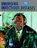
Edvard Munch (1863-1944). Self-Portrait After the Spanish Flu (1919-20)
| EID | Potter P. Edvard Munch (1863-1944). Self-Portrait After the Spanish Flu (1919-20). Emerg Infect Dis. 2003;9(3):407. https://doi.org/10.3201/eid0903.ac0903 |
|---|---|
| AMA | Potter P. Edvard Munch (1863-1944). Self-Portrait After the Spanish Flu (1919-20). Emerging Infectious Diseases. 2003;9(3):407. doi:10.3201/eid0903.ac0903. |
| APA | Potter, P. (2003). Edvard Munch (1863-1944). Self-Portrait After the Spanish Flu (1919-20). Emerging Infectious Diseases, 9(3), 407. https://doi.org/10.3201/eid0903.ac0903. |


